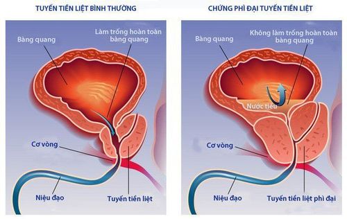
This is an automatically translated article.
The article is professionally consulted by Master, Doctor Nguyen Van Huong - Department of Diagnostic Imaging - Vinmec Danang International General Hospital.
Diagnostic method for benign prostatic hypertrophy, transrectal ultrasound can give the clearest picture, approach to accurately assess the size, as well as the lesion morphology, parenchyma and a number of lesions around the prostate gland.
1. What is benign prostatic hypertrophy?
Benign prostatic hypertrophy (or prostatic hyperplasia) is a benign enlargement of the prostate gland, the cause of which is due to the hyperplasia of the structural cells of the prostate gland, causing prostatic hyperplasia. symptoms of urinary disorders, early detection and treatment will avoid complications. Benign prostatic hypertrophy can develop slowly over a long period of time without causing any danger. However, because the gland surrounds the urethra, if the prostate is enlarged, it will cause some complications such as:
Complete urinary retention or not: Symptoms of urinary retention may occur after a long time when the patient Urinary incontinence may also have a sudden onset after a long latent period. Patients with symptoms feel a blockage in the urinary tract, it is difficult to urinate until complete urinary retention. In the complicated stage, the patient may fall into the state of urinary incontinence, frequent overflow of urine. Urinary tract infections: The stagnation of urine in the bladder creates conditions for bacteria to grow, causing many diseases such as cystitis, orchitis, prostatitis, ... Besides, the Urine reflux into the kidney can cause nephritis, retrograde pyelonephritis, more seriously, it can lead to sepsis, which is life-threatening. Bladder damage: Due to urinary disorders, the bladder has to endure being filled with urine for a long time, leading to the weakening of the bladder muscles.
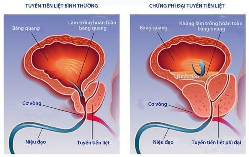
Phì đại lành tính tiền liệt tuyến
Kidney damage: Long-term urine stagnation will affect kidney function, reduce kidney function and will gradually progress to kidney failure. Blood in the urine: Blood in the urine is less common, usually at the top of the yard, but there are also cases of blood in the urine and blood clots. Benign prostatic hypertrophy also severely affects the psychological and sexual health of women due to experiencing painful symptoms during sex or increasing the risk of infertility. In addition, benign prostatic hypertrophy also causes a number of systemic diseases, such as cardiovascular disease, hypertension, trauma,...
2. Diagnosis of benign prostatic hypertrophy on transrectal ultrasound
Method of diagnosing benign prostatic hypertrophy, transrectal ultrasound can give the clearest picture, approach to accurately assess parenchyma, lesion morphology, size as well as surrounding lesions. Prostate.
2.1. Indications and contraindications
The method of diagnosing benign prostatic hypertrophy by rectal ultrasound is indicated when:
The patient has abnormal symptoms of urination. When transabdominal ultrasound is abnormal PSA test > 10ng/l and rectal examination palpable solid lesion. The method of diagnosing benign prostatic hypertrophy by rectal ultrasound is contraindicated when:
In case of severe infection Coagulation disorder, body exhaustion... Cardiovascular, respiratory diseases, Internal hemorrhoids Rectal narrowing due to tumor or infection.
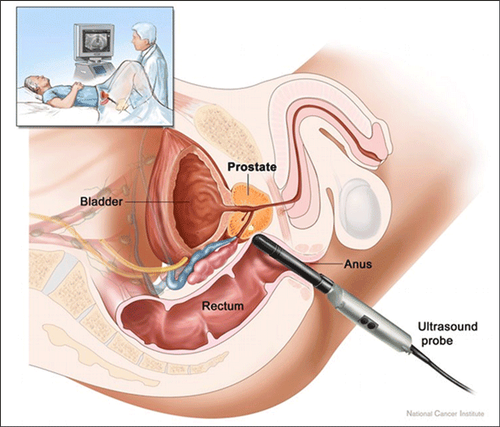
Phương pháp chẩn đoán phì đại lành tính tiền liệt tuyến qua siêu âm trực tràng
2.2. Steps to perform
Steps to perform transrectal ultrasound procedure to diagnose benign prostatic hypertrophy include:
Step 1: The doctor cleans the patient's rectum before performing the ultrasound. Mix 2 liters of water with Fortran and drink within 2 hours. Step 2: Ask the patient to lie on his side with his back to the doctor, bend his legs to bring his knees close to his chest. Then, the doctor examines and evaluates the anal area and surrounding soft tissue... Step 3: Use a condom and apply a layer of gel to the transducer to help conduct the sound outside. Then insert the probe slowly through the anus with light force. Step 4: After inserting the transducer through the anal sphincter and obtaining an image of the prostate, the sonographer selects the transducer with light force to get the best and clear picture of the prostate gland. . Step 5: When in doubt, a biopsy should be performed under the guidance of transrectal ultrasound, if there are nodules or suspected cancerous lesions, an additional biopsy sample should be taken at the same location. Step 6: Put the biopsy sample in the vials containing the specimen holding solution and number the sampling locations on each vial. Send the biopsy sample to the Pathology Department for necessary tests.
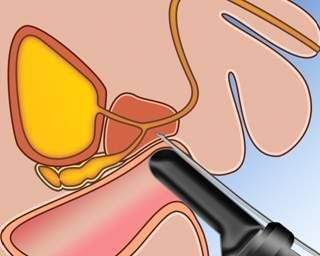
Sinh thiết phì đại lành tính tiền liệt tuyến
With his dedication to his work, dedication to the medical profession in general and the specialty of diagnostic imaging in particular, Dr. Huong is always loved and trusted by patients.
To register for examination and treatment at Vinmec International General Hospital, you can contact Vinmec Health System nationwide, or register online HERE.
Recommended video:
Close-up of prostate cancer treatment with robotic laparoscopic surgery
MORE:
Treatment of prostate enlargement with Bipolar knife - an advanced method to treat prostate cancer discharged from hospital 3 days after prostate surgery


