This is an automatically translated article.
The article is professionally consulted by Master, Doctor Tran Thi Vuong - Doctor of Microbiology - Laboratory Department - Vinmec Hai Phong International General HospitalInfection with tapeworm larvae is very dangerous, so it is very important to diagnose for appropriate and timely treatment. The diagnosis of cysticercosis should be confirmed by looking for the larvae in tissue biopsy specimens of the skin or subcutaneous tissue. In addition, the patient should be examined and palpated to find pea-sized nodules. Cysticercosis tests for tapeworm larvae are indicated as simple X-ray, CT scan, MRI,...
1. How dangerous are swine fluke larvae?
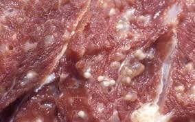
Tuberculosis is a parasitic disease capable of infecting animals and humans. The cause of the disease is mainly due to eating pork carrying tapeworm larvae or eating some foods contaminated with larvae in soil, water...Location of tapeworm larvae in common order is in CNS, subcutaneous tissue and skeletal muscle, and finally the eyeball, rarer than other organs. Protoscolex head, with 4 suction cups and rows of hooks, coils inside and attaches to the inner wall of the cocoon. Common locations and manifestations of infection with tapeworm larvae are as follows:
Nervous system In the nervous system, when rice fluke larvae develop here, it will manifest with symptoms of space-occupying, inflammatory reactions. or occlusion of the ventricular system and brain ducts. In the acute stage of invasion, the tapeworm larvae will cause a series of symptoms such as fever, headache, myalgia, eosinophilia, and worse, the patient falls into a coma.
Parenchymal cocoon presents with focal or generalized epileptic symptoms accompanied by focal neurological signs, increased intracranial pressure, and impaired consciousness. If the cyst is in the subarachnoid space and meningeal cocoon, it often leads to arachnoiditis, thereby causing increased intracranial pressure, hydrocephalus due to cerebral embolism, transient ischemia or infarction as well as dysfunction. cranial nerves.
The ventriculomegaly can float freely in the catheters of the brain or the ventricles, and in some cases it can attach to the wall of the ventricles. These cysts usually do not cause any symptoms but can also cause intracranial pressure due to total or intermittent occlusion.
Swine fluke larva is a rare abnormality. This larva has many branches, neither cocoons nor fluke head. They often form in unequal clusters, located in the subarachnoid spaces at the base of the skull and the ventricles, thereby causing arachnoiditis and obstructive hydrocephalus.
Spinal cord larva can appear externally or intramedullary, it often causes arachnoiditis such as meningitis, radiculopathy or symptoms of hypertension.
Eye fluke larvae In the case of ocular tapeworm larvae, they usually have only a free-swimming cocoon in the vitreous or subretinal. Symptoms of tapeworm infestation in the eye include pain around the eye, blurred vision, and blurred vision. Besides, some other clinical signs may appear, including: hemorrhage and edema of the optic disc, retinal detachment, iritis and retinitis.
Subcutaneous and striated muscle infection Under the skin and striated muscle, the subcutaneous larval cysts show signs of the appearance of nodules, which may collapse and disappear but then reappear. appear in different locations for different periods of time. These cysts usually cause no symptoms to the patient.
2. Cysticercosis test for tapeworm larvae
Infection with tapeworm larvae is very dangerous, so it is very important to diagnose for appropriate and timely treatment. The diagnosis of cysticercosis should be confirmed by looking for the larvae in tissue biopsy specimens of the skin or subcutaneous tissue. In addition, the patient should be examined and palpated to find pea-sized nodules. Cysticercosis tests for tapeworm larvae include:Muscle radiography alone: This imaging technique can detect oval or straight calcified lesions of tapeworm larvae. These lesions are often numerous and are located along the direction of the surrounding muscle fibers. CT scan: One of the most effective brain exploration is diagnostic CT Scan. When using this technique, the images of parenchymal cysts on the film will show water cysts (areas of circular hypodense that do not capture contrast); glue cocoons are dead or dying cocoons that have the body's immune response; granuloma or calcification. CT Scan can detect calcified lesions and granulomas. Magnetic resonance imaging (MRI): This technique gives a clearer image of the water cocoon and the glue cocoon. However, the specific nodules 2 - 4 mm in the cocoon fluid are less specific. Cases of cysticercosis in the spinal cord can be detected by magnetic resonance imaging (MRI). Immunoassays: One test with 100% specificity and 95% sensitivity for Cysticercosis is the new serum immunoelectrophoresis fixation test. Another test method is the ELISA method with a specificity of 63% and a sensitivity of 65%. Cerebrospinal fluid test: With this Cysticercosis test technique, sensitivity is up to 86% with immunofixation response. Cerebrospinal fluid in neurocysticercosis should be tested for IgM and IgG antibodies; cerebrospinal fluid often increases protein but decreases glucose; Eosinophilia more than 20% is also valuable in the diagnosis of tapeworm larvae.

3. Treatment and prevention of tapeworm larvae
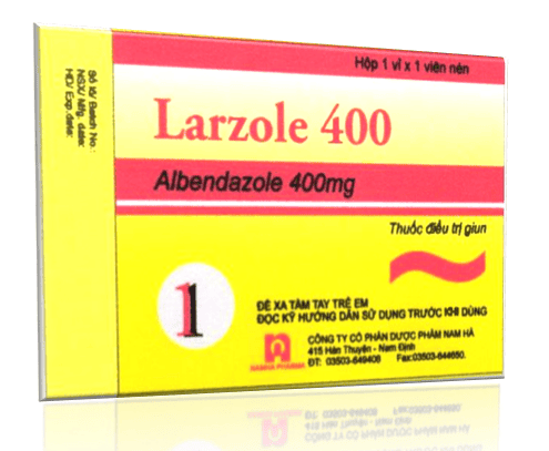
3.1. Treatment of swine flu When the infection is detected with tapeworm larvae, the patient needs medical treatment to destroy the tapeworm, in some cases, surgery is required. However, the treatment of tapeworm larvae must be carried out in medical facilities with modern equipment and experienced medical staff. Treatment methods for tapeworm larvae include:Drug treatment: The drugs used to treat tapeworm larvae are Praziquantel, Niclosamide and Albendazole. In the case of tapeworm larvae causing nerve compression or causing embolism, ventricular dilatation or water retention in the brain, the patient needs to perform surgical methods as quickly as possible. 3.2. Prevention of tapeworm larvae To actively prevent infection with tapeworm larvae, people need to keep the environment clean and manage waste properly. Besides, people need:
Use clean water in all daily activities. Wash your hands with soap under running water before and after using the toilet, before cooking and eating, and wash your body often. Eat cooked and drink boiling; choose reputable pork origin, do not eat pork and products processed from pork when it is not cooked. At the same time limit the consumption of raw vegetables as much as possible. Sanitary slaughter of pigs, especially do not let pigs loose. Small fluke disease, if not detected early and treated promptly, will seriously affect the patient's health. Therefore, as soon as signs and symptoms of the disease appear, people need to go to medical centers for timely examination and treatment.
Currently, Vinmec International General Hospital is one of the leading prestigious hospitals in the country, trusted by a large number of patients for medical examination and treatment. Not only the physical system, modern equipment: 6 ultrasound rooms, 4 DR X-ray rooms (1 full-axis machine, 1 light machine, 1 general machine and 1 mammography machine) , 2 DR portable X-ray machines, 2 multi-row CT scanner rooms (1 128 rows and 1 16 arrays), 2 Magnetic resonance imaging rooms (1 3 Tesla and 1 1.5 Tesla), 1 room for 2 level interventional angiography and 1 room for bone mineral density measurement... Vinmec is also the place to gather a team of experienced doctors and nurses who will greatly assist in diagnosis and early detection. abnormal signs of the patient's body. In particular, with a space designed according to 5-star hotel standards, Vinmec ensures to bring the patient the most comfort, friendliness and peace of mind.
Please dial HOTLINE for more information or register for an appointment HERE. Download MyVinmec app to make appointments faster and to manage your bookings easily.


