This is an automatically translated article.
The article was professionally consulted by Specialist Doctor I Nguyen Truong Duc - Radiologist - Department of Diagnostic Imaging and Nuclear Medicine - Vinmec Times City International General Hospital.Coronary calcification is a warning sign of coronary artery disease. Computed tomography (CT) assessment of coronary calcification is a modern technique that allows to detect calcifications on the coronary artery wall. The CT scan procedure to evaluate coronary calcification is simple, non-contrast and non-invasive.
1. What is a CT scan to assess coronary calcification?
Computed tomography assessment of coronary calcification, also known as CT scan for calcification, has the English nomenclature as Heart scan or coronary calcium scan or Coronary artery calcium (CAC) scoring is an angiographic technique. Coronary by computer tomography machine but not iodized contrast medium for the purpose of detecting calcification points on the coronary system.By imaging the calcification points on the coronary artery system, this technique allows to assess the degree of calcification of the coronary arteries.
Coronary calcification is a warning sign of coronary artery disease. Therefore, CT scan to evaluate coronary calcification allows to predict problems, cardiovascular disease, heart attack before symptoms caused by disease.
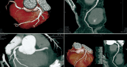
Chụp CT scan giúp chẩn đoán, đánh giá tình trạng vôi hóa mạch vành
2. Indication for CT scan to evaluate coronary calcification
CT scan to assess coronary calcification is indicated in the case of screening for coronary artery calcification in patients with risk factors for cardiovascular disease such as:Hypertension
● Blood lipids high
● Smoker
● A family member with cardiovascular disease
● Diabetic patient.
● Obesity.
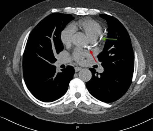
Coronary calcification is a warning sign of atherosclerosis causing a heart attack

Vôi hóa mạch vành là dấu hiệu cảnh báo xơ vữa động mạch gây đau tim
3. CT scan procedure to evaluate coronary calcification
Patients should be careful not to smoke or drink coffee about 4 hours before the scan. The procedure of CT scan to evaluate coronary calcification includes the following steps:● Step 1: Remove the necklace, personal clothing and put on a medical gown. Be explained and guided by medical staff to coordinate well during the imaging process. Place the patient supine on the imaging table.
● Step 2: The technician glues the electrodes and installs the electrocardiogram port, checking the patient's heart rate on the screen of the scanner.
● Step 3: Take coronary angiography with the field from the bronchial fork to the end of the apex, the thickness of the cut layer is 3mm.
● Step 4: The technician reproduces the image on the camera at the shooting room. The amount of calcium detected in the coronary artery wall and assessed by the Agatston scale will give the corresponding coronary calcification results. The result is negative if the scanner does not detect calcification on the coronary artery wall and the patient is at low risk of heart attack. Conversely, the result is positive if the scanner detects calcifications on the coronary walls. The higher the results, the higher the risk of developing atherosclerosis and leading to heart attacks.
CT scan to evaluate coronary calcification is a non-invasive technique, without using contrast and the risk of radiation contamination is very low. Therefore, this is a safe technique to help detect and evaluate calcified coronary arteries, preventing atherosclerosis causing heart attacks.
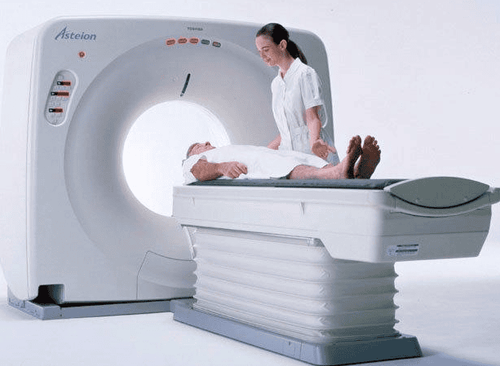
Chụp CT đánh giá vôi hóa mạch vành là kỹ thuật tiên tiến đã được áp dụng tại Bệnh viện Đa khoa Quốc tế Vinmec
Doctor Duc has nearly 17 years of experience in diagnostic imaging at Hanoi E hospital. Currently, the doctor is working at the Department of Diagnostic Imaging - Vinmec Times City International Hospital.
To register for examination and treatment at Vinmec International General Hospital, you can contact the nationwide Vinmec Health System Hotline, or register online HERE.