This is an automatically translated article.
The article was professionally consulted by resident Doctor Nguyen Quynh Giang - Department of Diagnostic Imaging and Nuclear Medicine - Vinmec Times City International Hospital.Coronary MRI is a high-tech imaging method that not all medical facilities can perform. The purpose of coronary magnetic resonance angiography is to evaluate atherosclerotic lesions, stenosis, or anatomical abnormalities of the coronary arteries.
1. What is coronary magnetic resonance angiography?
Coronary artery MRI is an imaging method with high spatial resolution, helping to accurately evaluate coronary artery diseases such as coronary occlusion, congenital abnormalities..., and preoperative assessment. and follow-up after treatment.Coronary magnetic resonance angiography also allows to further evaluate the morphology and movement of 2 or 4 chambers of the heart in coronary artery disease (myocardial ischemia), congenital heart disease ....
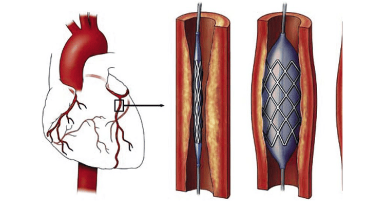
Bệnh nhân mắc động mạch vành có thể gây ra nhiều biến chứng nguy hiểm về sức khỏe
2. Indications for coronary artery MRI
Indication for coronary MRI when:● When the patient has symptoms of chest pain, shortness of breath, dizziness...then the doctor may order the patient to have coronary MRI to evaluate the symptoms. anatomical abnormalities.
● Survey of coronary artery disease : coronary aneurysm in Kawasaki disease ...
Preoperative assessment for congenital heart disease .
3. Contraindications
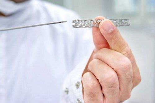
● Patients wear electronic devices such as: cochlear implants, defibrillators, pacemakers, Neurostimulators, automatic drug injection devices under the skin....● Wear metal surgical clips Intracranial, vascular, orbital for less than 6 months (If worn for more than 6 months, consider further depending on the patient's health status).
● People with serious illnesses need resuscitation equipment on their side.
People with kidney failure, severe liver failure (consider)
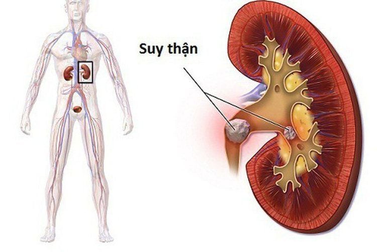
Bệnh nhân suy thận chống chỉ định thực hiện
4. Coronary magnetic resonance imaging procedure
4.1 Preparation for the procedure
To perform coronary magnetic resonance angiography, it is necessary to prepare:Implementation team:
● Radiology specialist.
Electro-optical technician.
Vehicles used:
Resonant angiography machine from 1.5 Tesla or more.
● Image storage system.
Film printer.
Movies.
Supplies used:
● 18G vein puncture needle.
● 10ml syringe.
● Physiological saline / distilled water.
● Gauze, gloves, sterile bandages.
● Contrast drug accident first aid box.
Drugs:
● Magnetic Contrast Drugs.
Sedatives.
Antiseptic of mucous membranes and skin.
Thuốc an thần có thể sử dụng nếu người bệnh quá kích động trước khi chụp
● The procedure is explained in advance to coordinate well with the doctor.
● Prepare a copy of the request from a clinician, with a clear diagnosis or complete medical record (if necessary).
● Check for contraindications.
● Change into specialized clothes of the magnetic resonance imaging room according to instructions, remove contraindicated items.
4.2 Implementation Procedure
The whole procedure of coronary MRI takes about 30 minutes in turn, following these steps:Patient position:
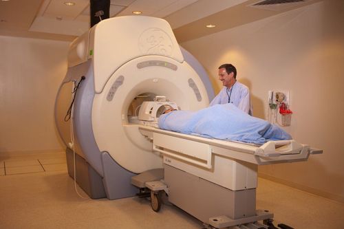
● The patient is instructed to lie supine on the scanning table.
● Insert the electrocardiogram electrode.
● Select and position the receiver coil.
● Move the table into the machine bay and locate the area to be photographed.
Shooting techniques
● Positioning shooting.
● Depending on the pathology and clinical requirements, the technician conducts the scan according to the appropriate procedures.
● Take pulse sequences to evaluate morphology and function on the planes of 2 chambers, 4 chambers, short axis, right ventricular outflow tract, left ventricular outflow...
● Coronary pulse sequence imaging (Whole heart 3D) ), which captures the signal according to the patient's breathing rate.
● Inject intravenous contrast to the patient to show the coronary artery lumen. The usual dosage is 0.1mmol/kg body weight.
Note: In the process of taking contrast agents, allergic reactions (itching, hives, nausea, anaphylaxis) may occur, so it is necessary to prepare for an emergency in case of an accident. .
● The technician processes the acquired images, prints the film, and transfers the images and data to the doctor's workstation. The image must clearly show the anatomical structure of the coronary arteries. Based on the above information, the doctor analyzes the lesion (if any) and makes a diagnosis.
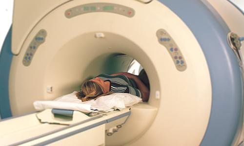
Khi chụp MRI động mạch vành, bệnh nhân nằm ngửa trên bàn chụp
5. Benefits of Coronary Magnetic Resonance Imaging
Coronary MRI is a modern imaging technique in medicine that uses magnetic fields to evaluate atherosclerotic lesions, stenosis or anatomical abnormalities of the coronary arteries. Images from MRI have high contrast, good anatomical detail, allowing accurate detection of lesions in terms of morphology, structure of parts in the body.Doctor Giang has many years of experience in the field of diagnostic imaging, especially in the field of multi-slice computed tomography, magnetic resonance. Currently, the doctor is working at the Department of Diagnostic Imaging and Nuclear Medicine - Vinmec Times City International General Hospital.
For medical examination and treatment at Vinmec, please go directly to Vinmec medical system nationwide or register online HERE.


