This is an automatically translated article.
The article was written by Doctor Le Van Binh - Department of Intensive Care - Vinmec Times City International Hospital.Congenital heart disease is the most common malformation and the leading cause of death among birth defects in children. Congenital heart disease is divided into cyanotic congenital heart disease and non-cyanotic congenital heart disease.
I- GENERAL
Congenital heart disease is an abnormality in the structure of the heart and large blood vessels, the disease is present at birth, the disease process parallels the development of the fetus
General frequency of congenital heart disease in the world About 8/1000 live births, the structure of the disease is quite complicated and early death.
There are many causes that affect or cause congenital heart disease, pathogenesis factors affecting the development of the fetal cardiovascular system such as: bacterial infection, viral infection (especially Rubella virus, influenza virus), chemistry poisons, X-rays, radiation, genetic inheritance (<10%), family factors (2-5%), or use of drugs such as pain relievers, antibiotics, antiepileptics, hormones..
Current diagnosis of congenital heart disease is usually based on clinical, electrocardiogram, chest radiograph, cardiac catheterization angiography and echocardiography. Thanks to the advent of 2D echocardiography and color Doppler, today most cases of congenital heart disease have a definitive diagnosis and surgery is indicated after performing 2D echocardiography and color Doppler, without the need for information. angiographic heart. The advent of heart surgery has brought positive results in the treatment of congenital heart disease.

II. CLASSIFICATION (Clinically based)
Congenital heart disease is usually divided into cyanotic congenital heart disease and non-cyanotic congenital heart disease. There are many classifications based on clinical, anatomical, or embryology.
2.1- Congenital heart disease without cyanosis a. Abnormalities originating from the left side of the heart:
+ Obstruction of the left atrium entrance: including pulmonary venous stenosis, mitral stenosis, triatrial heart.
+ Mitral regurgitation
+ Primary endocardial elastosis
+ Aortic stenosis (subvalvular, valvular and supravalvular).
+ Aortic valve regurgitation.
+ Coarctation of the aorta.
b. Abnormalities originating on the right side of the heart
+ Ebstein's disease
+ Pulmonary stenosis (under the funnel, funnel, valve, and above the valve).
+ Congenital pulmonary valve regurgitation.
+ Idiopathic pulmonary artery dilatation.
+ Primary pulmonary hypertension.
2.2. Congenital heart disease without cyanosis a. Atrial septal flow
+ Atrial septal defect (1st hole, 2nd hole, venous sinus, coronary sinus).
+ Abnormal connection of partial pulmonary veins.
+ Atrial septal defect with mitral stenosis (Lutembacher syndrome).
b. Ventricular flow
+ Ventricular septal defect (perimembrane, funnel area, receiving chamber, trabecular region).
+ Ventricular septal defect with aortic regurgitation).
+ Ventricular septal defect has left atrial septal flow.

+ Fistula of the coronary artery.
+ Valsalva sinus aneurysm rupture.
+ The left coronary artery originates from the pulmonary artery.
d. Flow between the aorta and the pulmonary artery.
+ The master window.
+ Have ductus arteriosus.
e. Superior Flow I Floor
+ Atrioventricular Channel
III. SOME CONTINUOUS HEART DISEASES WITHOUT CLEANING WITHOUT TRAFFIC.
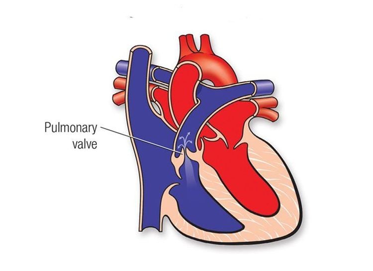
3.1. Pulmonic stenosis (PS) * General:Pulmonary stenosis can be caused by damage to the pulmonary valve (pulmonary valve stenosis), damage to the funnel (exit stenosis) due to hypertrophy of the supraventricular muscle Ventricular stenosis or stenosis combines valve and funnel while the interventricular septum remains normal. Pulmonary stenosis may be isolated, possibly in the setting of tetralogy of Fallot.
* Pathophysiology:
Right ventricle has blood stasis, increased load and can lead to right ventricular failure. Cardiac output decreases due to decreased left ventricular blood flow from the lungs.

* Diagnostic symptoms:
+ Systolic blow in the pulmonary valve.
+ Electrocardiogram: right ventricular thickening.
+ Cardiac catheterization: right ventricular pressure increases while pulmonary pressure decreases.
+ Ultrasonography: Determine the stenosis, pressure difference between the right ventricle and the pulmonary artery.
* Surgery:
+ Indications: Pulmonary stenosis with pressure difference between right ventricle and pulmonary artery over 50 mmHg, appropriate age for surgery over 5 years old.
+ Methods:
- If the pulmonary valve stenosis is simple: wide dissection of the orifice can be performed under temporary circulatory arrest (temporary clamping of 2 vena cava) or under an artificial cardiopulmonary machine.
- If narrowing of the funnel or stenosis of the valve-funnel mixture: surgery must be performed under an artificial heart-lung machine. Open the ventricle to remove the area causing the stenosis, if the stenosis remains after resection, it may need to be patched to widen the area.
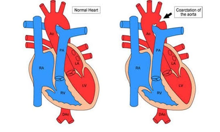
3.2. Coarctation of the aorta (CoA). * Outline:
Aortic stenosis can occur at different locations, but the most common is isthmus aortic stenosis (the area corresponding to the aorta-pulmonary ligament, behind the separation of the left subclavian artery).
* Pathophysiology:
Blood stagnates on the stenosis, which increases blood pressure in the arteries in the upper extremities and skull, but causes ischemia in the lower part of the body. The left heart has to increase the force of contraction, so it is often overloaded.
* Diagnostic symptoms:
+ Unbalanced body development; upper body (2 upper limbs, large neck while 2 lower limbs are small and slender). The blood pressure in the arms is high while the blood pressure in the legs decreases (normally, the systolic blood pressure in the legs is about 10-20 mmHg higher than in the arms).
+ A systolic murmur can be heard at the left edge of the sternum, the fourth and fifth intercostal spaces.
+ Electrocardiogram: there is often increased left ventricular load.
+ X-ray: The lower border of the ribs has a V-shaped concave slit due to the dilated intercostal arteries, the left ventricle is enlarged.
+ Ultrasound: can identify aortic stenosis.
+ Contrast aorta: accurately identify aortic stenosis as well as its lateral circulation.

* Surgery:
+ Indications: All patients who can tolerate surgery should have surgery, the most suitable age for surgery is 7-10 years old.
+ Surgical method:
Bypass through the stenosis: usually a piece of artificial blood vessel is used to bridge the bridge between the upper and lower parts of the stenosis. Vascular patching: cutting along the artery wall in the narrow area and using a piece of patch material to widen the artery. End-to-end anastomosis after resection of the stenosis (only used when the excised stenosis is not too long). Vasectomy: Using an artificial artery to graft after resection of the narrow artery. Recently, the method of percutaneous narrowing artery dilation is applied. Technique: insert a percutaneous arterial catheter, bring the balloon to the stenosis, and inflate the balloon to widen the lumen. + Early complications after surgery: high blood pressure, abdominal pain, paralysis of the lower extremities and chylous pleural effusion (Chylothorax). Distant complications: aortic aneurysm, restenosis.
Vinmec International General Hospital is a high-quality medical facility in Vietnam with a team of highly qualified medical professionals, well-trained, domestic and foreign, and experienced.
A system of modern and advanced medical equipment, possessing many of the best machines in the world, helping to detect many difficult and dangerous diseases in a short time, supporting the diagnosis and treatment of doctors the most effective. The hospital space is designed according to 5-star hotel standards, giving patients comfort, friendliness and peace of mind.
If you have a need for consultation and examination at Vinmec Hospitals under the national health system, please book an appointment on the website for service.
Please dial HOTLINE for more information or register for an appointment HERE. Download MyVinmec app to make appointments faster and to manage your bookings easily.


