This is an automatically translated article.
The article is expertly consulted by Master, Doctor Luu Thi Bich Ngoc - Doctor of Radiology - Department of Diagnostic Imaging and Nuclear Medicine - Vinmec Times City International General Hospital.Some acute diseases such as aortic dissection, if not diagnosed and treated promptly, have a very high risk of death. The method of computed tomography of the aorta helps to quickly and accurately diagnose some acute and chronic diseases of the aorta.
1. What is computed tomography of the thoracic aorta?
Computed tomography of the thoracic aorta is a method of using an X-ray machine to project the entire thoracic aorta in slices, then the machine will process the images by computer, then the doctor will aim to examine abnormalities of the thoracic aorta.2. Indications and contraindications of thoracic aorta computed tomography
Indications:● Chest pain suspected of acute aortic syndrome such as: aortic dissection, arterial thrombosis, intra-wall hematoma or trans-aortic ulcer.
● Suspected case of coarctation of the aorta.
● Inflammatory diseases of the aorta: Takayasu...
● Assess the status of atherosclerosis of the aortic wall and major vessel roots arising from the aortic arch causing narrowing of the vessel lumen
Cases of trauma thoracic area suspected thoracic aorta injury.
Contraindications:
● Relative contraindications: Pregnant women, patients who do not agree to take pictures.
● Absolutely contraindicated in cases with contraindications to iodine contrast injection such as: Patients with severe renal failure, liver failure, severe heart failure, history of contrast allergy, chronic disease diabetes, bronchial asthma, untreated hyperthyroidism, sickle cell anemia...
3. Computerized tomography of the thoracic aorta
3.1 Preparation
Performer: Team of doctors and technicians specializing in diagnostic imaging.Means used to take computed tomography include:
● Multi-segment computed tomography machine, specialized electric injection pump.
● Film capture, photo printer and image storage system.
Water-soluble iodine contrast agent.
● Medical supplies include: Syringes and needles (type 10ml, 20ml, 50ml), syringes for electric syringes, threaded needles (18-20G), antiseptic solution, physiological saline, gloves hand, mask, tray set, cotton gauze, bean tray, surgical gauze, surgical forceps.
● Anti-shock box, drugs and necessary tools to handle accidents in case of abnormality when the patient injects contrast agent.
Patients:
● Patients are clearly explained about how to take pictures and possible complications when taking pictures to coordinate with the photographer.
● Remove items that can cause image noise and affect image quality such as necklaces, bras... (if any).
● You need to fast for at least 4-6 hours before the scan. You can drink water but not more than 50ml.
● The patient is overstimulated, anxious and afraid or uncoordinated when taking the picture: It is necessary to give sedation as prescribed by the treating doctor before taking the picture.
3.2 Steps to take
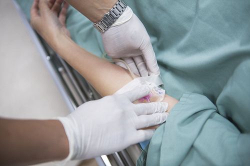
● Patient position: Place the patient in supine position, place a large enough intravenous line and connect to an electric syringe. The ECG port may or may not be fitted.● Positioning scan: Carry out a positioning scan from the neck to the level of the diaphragm, making sure to get all of the origin and proximal part of the great vessels originating from the aortic arch.
● Pre-injection imaging: Usually only taking the chest segment, evaluating the hematoma lesions in the wall with natural increase in density.
● Taken after contrast injection with a slice thickness corresponding to the pre-injection phase:
● After the image is reconstructed, the doctor will read the results and make a judgment.
3.3 Evaluation of results
● The layers are symmetrical, the image is clear, the thoracic aorta is clearly distinguished on the reconstructed images.● See abnormal changes in the thoracic aorta if present.
● The doctor reads and describes the lesion, then prints the results.
4. Possible complications with computed tomography of the thoracic aorta
Possible complications due to contrast media:● Anaphylaxis
● Contrast-induced renal failure
● Thyroid storm: Manifestations of increased temperature, tachycardia, emotional disturbances, otherwise If detected signs of worsening pulmonary edema, heart failure, coma...
● Pregnant women need to consider carefully before taking the picture and when taking the picture, they must wear a lead shirt to protect the abdomen.
Master, Doctor Luu Thi Bich Ngoc has 06 years of experience working in the field of diagnostic imaging, received formal training and graduated with a Master's degree from Hanoi Medical University. Dr. Ngoc has strengths in diagnosing breast, thyroid, liver cancers early. experience in doing ultrasound of tissue elastography for liver, breast, thyroid diseases... From 2019 until now, Dr. Ngoc has worked at Vinmec Times City International Hospital with a position at the department. Image analysation.
To register for examination and treatment at Vinmec International General Hospital, you can contact Vinmec Health System nationwide, or register online HERE.
MORE
Magnetic resonance imaging (MRI) of the thoracic aorta: What you need to know Magnetic resonance imaging (MRI) of the thoracic aorta (thoracic-abdominal segment) Thoracic magnetic resonance imaging (MRI) procedure


