This is an automatically translated article.
The article is professionally consulted by MSc, BS. Dang Manh Cuong - Doctor of Radiology - Department of Diagnostic Imaging - Vinmec Central Park International General Hospital. The doctor has over 18 years of experience in the field of ultrasound - diagnostic imaging.Contrast-injected cranial computed tomography is an advanced imaging technique, widely applied in the diagnosis of neurological diseases. In which, cranial CT scan is very effective in assessing the degree of angiogenesis of tumors, the extent of lesions such as encephalitis, meningitis, inflammation of the brain parenchyma.
Meningitis patients in the process of diagnosis and treatment will be assigned a CT scan of the brain with contrast injection to increase the diagnostic efficiency and assess the level of treatment.
1. What is meningitis?
Meningitis is an inflammation of the thin membrane covering the brain and spinal nerves. Meningitis can be caused by bacteria, viruses from elsewhere in the body that spread through the bloodstream into the cerebrospinal fluid, or in rare cases, fungi, parasites, or reactions to chemicals. substances, autoimmune diseases.Up to 25% of patients with meningitis have symptoms that progress rapidly within 24 hours, the rest develop in 1-7 days. It can be recognized by typical signs such as headache, stiff neck, fever, chills, vomiting, photophobia, convulsions and symptoms of upper respiratory tract infection. In addition, the patient may have atypical symptoms such as focal weakness, reduced sensation of movement, pain and swelling of one or more joints, a rash that looks like a bruise...
When having the above symptoms, Patients need to be examined at reputable and qualified medical facilities. As directed by a specialist, the patient will be assigned a CT scan of the brain with contrast injection to diagnose meningitis accurately and promptly.
Some cases of patients contraindicated to CT cranial contrast injection are:
In the examined brain area, there are many metals that interfere with the image. Cases of contraindication to contrast injection Some cases In case of relative contraindications such as pregnant women, especially in the first 3 months of pregnancy, when taking pictures, it is necessary to use a lead jacket to cover the abdomen.
2.Preparing for a CT scan of the brain with contrast injection
Consumables include:
Syringe and syringe 10, 20ml, 50ml Needle conduit 18-21G Water-soluble iodine contrast solution Skin and mucosal antiseptic solution Distilled water or physiological saline Gloves, caps, Surgical mask Set of bean trays, surgical forceps Surgical cotton and gauze Medicine box, emergency equipment for contrast drug accidents Patients with CT scan of the brain injecting contrast are explained in detail about the technique to coordinate shooting. Family members or patients sign a commitment to perform the technique. The patient must remove the earrings, necklace, and hairpins before taking the picture. Besides, need to fast, drink before 4 hours, can drink but not more than 50ml of water. For patients who are overstimulated, do not lie still for the scan, need to give sedation as prescribed by the treating doctor before the scan.
3. Conducting a CT scan of the brain with contrast injection

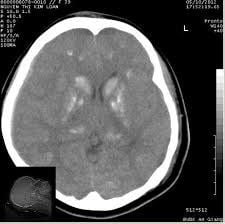
Symmetric tomography Good image contrast, suitable for distinguishing white matter, gray matter Showing abnormal changes in density, morphology of brain, meninges, bones, sinuses, software before and after contrast injection The specialist reads the lesions, describes on the computer and prints the results. Professional advice can be given to the patient and family if required
4.Complications and treatment
The patient may be frightened, agitated, in need of encouragement, or sedated. Some cases of complications with contrast agents need to be handled quickly according to the procedure.The CT scan of the brain with contrast injection helps in the early and effective diagnosis of meningitis. Patients with abnormal symptoms mentioned above should go to the doctor and implement the earliest methods of diagnosis and treatment.
Vinmec International General Hospital is a high-quality medical facility in Vietnam with a team of highly qualified medical professionals, well-trained, domestic and foreign, and experienced.
A system of modern and advanced medical equipment, possessing many of the best machines in the world, helping to detect many difficult and dangerous diseases in a short time, supporting the diagnosis and treatment of doctors the most effective. The hospital space is designed according to 5-star hotel standards, giving patients comfort, friendliness and peace of mind.
Please dial HOTLINE for more information or register for an appointment HERE. Download MyVinmec app to make appointments faster and to manage your bookings easily.


