This is an automatically translated article.
The article is professionally consulted by Doctor CK II Nguyen Quoc Viet and Master, Doctor Pham Van Hung - Department of Medical Examination & Internal Medicine - Vinmec Danang International General HospitalRestrictive myocardiopathy is a disease with low morbidity but severe complications and high mortality. There are many causes of the disease, but the most common cause is primary endocardial fibrosis, if untreated, the patient can die after 2-3 years. Therefore, early detection and treatment of the disease can help prolong the patient's life.
1. What is restrictive cardiomyopathy?
Restrictive cardiomyopathy is the inability of the ventricles to relax enough to fill with blood (decreased diastolic function). The ventricles are the main chambers of the heart responsible for pumping blood to the lungs for oxygen exchange and for pumping oxygen-rich blood from the heart to the body.
When the ventricles are not filled with blood, it will reduce the amount of blood to nourish the organs, making the patient always feel tired, short of breath, even have the initial symptoms of heart failure.
Damage to diastolic function is initially the result of limited ventricular dilatation, and later due to ventricular chamber obstruction. In the late stages of heart failure, there may be pericardial effusion.
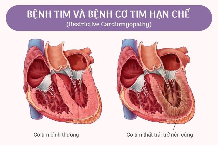
2. Causes of restrictive cardiomyopathy
Endomyocardial fibrosis is a common cause of restrictive cardiomyopathy; endocardial fibrosis of unknown cause predisposes to primary restrictive cardiomyopathy.In addition, other possible causes of the disease include:
Iron overload disease. Starch degenerative disease. Sarcoidosis (inflammation of the lymph nodes and tissues) Systemic scleroderma After radiation therapy or chemotherapy prior cancer treatment Rejection following a heart transplant. Diseases of the endocardium.
3. Symptoms of restrictive cardiomyopathy
Symptoms of restrictive cardiomyopathy vary depending on the location of the ventricular damage, they may include:Dyspnea on exertion Precardiac pain, liver pain (easily confused with hepatobiliary disease) . Feeling of heart palpitations, fluttering in the chest (palpitations) The patient feels dizzy, tired, or faints when exercising, exerting himself or standing up suddenly while sitting. Feeling tired, not wanting to work. Nausea and anorexia Weight gain and swelling, edema in the legs, ankles, feet and abdomen bulging veins in the neck On examination, a systolic murmur can be heard due to 2-3 leaf regurgitation, palpable hepatomegaly.

4. How to diagnose disease?
In addition to relying on clinical signs, to diagnose the disease, it is necessary to perform laboratory tests including:Electrocardiogram (ECG): There is almost always an abnormal electrocardiogram. Left bundle branch block and atrial thickening are common findings. Cardiopulmonary : The heart shadow is usually not enlarged unless there is dilated atrial enlargement, pulmonary congestion is often severe. Echocardiography: Detects endocardial fibrosis. Assess ventricular status, heart function, valves and pericardium, detect mitral regurgitation and other valves, and diastolic dysfunction such as abnormalities such as dilation and dilation of the ventricles. Blood tests look for a number of causes, such as: Eosinophilia increased in endocardial fibrosis. Quantification of serum iron, ferritin (storage iron) to assess the status of iron excess, immune bilan (scleroderma). In addition, in some hospitals with full equipment, a number of diagnostic techniques can be used such as:
Computed tomography (CT) and magnetic resonance (MRI): Give images to help distinguish from constrictive pericarditis due to signs of pericardial thickening. Cardiac catheterization: Indicated in cases requiring differential diagnosis with constrictive pericarditis and also serves the purpose of myocardial biopsy to diagnose the cause of restrictive cardiomyopathy. Endomyocardial biopsy: Permits a definitive diagnosis and can lead to a diagnosis of etiology.
5. Disease progression and prognosis
The course of the disease depends on the cause of the disease:May progress quickly to heart failure with paroxysmal breathlessness, generalized edema, venous occlusion. Patients can also die suddenly due to severe arrhythmias, or experience thromboembolic complications due to the formation of blood clots, leading to stroke, myocardial infarction, kidney failure, hand necrosis. For idiopathic restrictive cardiomyopathy (endocardial fibrosis), if untreated, the patient can die after 2-3 years, if detected early, monitored and intervened promptly The 10-year survival rate is 50%. Restrictive cardiomyopathy is a dangerous disease with high mortality. Early detection and treatment help increase life expectancy and improve quality of life.
Please dial HOTLINE for more information or register for an appointment HERE. Download MyVinmec app to make appointments faster and to manage your bookings easily.


