This is an automatically translated article.
This article is professionally consulted by Master, Doctor Vu Huy Hoang - Radiologist - Department of Diagnostic Imaging and Nuclear Medicine - Vinmec Times City International Hospital.Tumor hemodynamic computed tomography is a modern technique that provides markers of angiogenesis and the state of blood vessels in the tumor.
1. In what cases is computed tomography tumor hemodynamic analysis (CT perfusion) indicated?
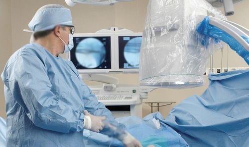
Computed tomography, also known as CT, is a technique that uses X-rays with cross-sections to examine parts of the body. The imaging system is linked with the computer, the algorithms and the computer processor help create 2D and 3D images for diagnosis.With the new generation of computed tomography machines, it can be easily integrated into the CT perfusion technique. This is a technique that helps to provide markers of angiogenesis in the tumor and reflect the status of blood vessels in the tumor. Tumor hemodynamic computed tomography scan helps doctors get more information about imaging in cancer pathology, not only useful in diagnosis, but also helps assess risk and monitor the following tumors. treatment.
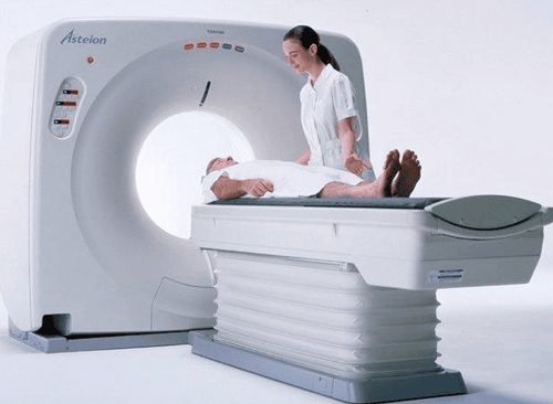
Chụp cắt lớp vi tính là kỹ thuật không xâm lấn, an toàn cho người bệnh
2. What do you need to prepare for a computed tomography scan for tumor hemodynamics?
Technical team includes specialist doctors, radiology technicians, nursesNecessary facilities include:
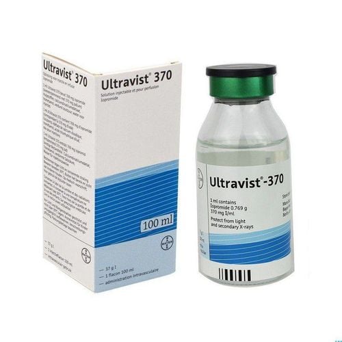
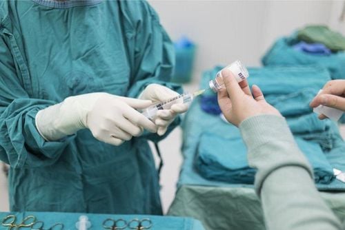
Computerized tomography machine Specialized electric pump Film, cassette (remove), image storage system In addition, to inject contrast, it is necessary to prepare medical supplies and drugs such as: syringe, needle, cotton, surgical gauze, contrast agent, skin antiseptic solution, water distillation, physiological saline, medicine box and emergency equipment for contrast dye accidents,...
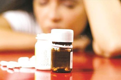
Before taking the picture, the patient will be thoroughly explained by the medical staff about the procedure, steps taken to coordinate with the physician. Remove jewelry such as necklaces, earrings and other metal objects on the body, if any. Instruct the patient to fast for at least an hour before the procedure. Small amounts of water can be drunk, no more than 40ml. If the patient is too excited to lie still, the doctor may prescribe a sedative to help the patient relax.

Thuốc an thần được chỉ định cho người bệnh trước khi chụp nhằm tránh kích động
3. Steps to conduct computed tomography of tumor hemodynamic studies
3.1. Prepare the patient
The doctor reviews the medical records, looks for signs of contraindications to intravenous iodinated contrast, and consults previous imaging results (if any). The nurse prepares the patient's intravenous line with an 18G needle.3.2. Steps to conduct tumor hemodynamic tomography (CT perfusion) technique
Instruct the patient to lie supine on the table, hands above the head. Take pre-injection slices of the entire abdomen (thorax, pelvis,...) depending on the tumor location. At the end of the shooting time, exhale. The doctor considers a preliminary assessment of the tumor in terms of location, density, size,... Then selects localized slices of about 2cm on the site with the largest diameter of the tumor. Make post-injection slices localized to the selected area, patient holds breath at the end of expiration, at a rate of 1 second for one cut, slice thickness 5-10mm, lasts about 25-30s in one session. breath hold. Contrast injection speed 6ml/s, drug dosage from 40-70ml depending on the examination department. After capturing, the image data will be transferred to a computer with measurement software, perfusion mapping, and enhancement chart. The doctor measures the most strongly enhanced tumor sites to compare with the enhancement chart of the aorta and the intact visceral parenchyma.
Hình ảnh chụp cắt lớp khảo sát huyết động khối u được xử lý qua phần mềm
In order to meet the needs of medical examination and treatment, currently Vinmec International General Hospital has been and continues to bring in a system of modern machines such as magnetic resonance imaging (MRI), computed tomography (CT), X-ray, etc. ... in the work of medical examination and treatment, diagnostic imaging, disease treatment. Especially in order to bring high efficiency in medical examination and treatment, Vinmec now also designs many accompanying medical services, bringing many conveniences to customers.
Doctor Vu Huy Hoang has 10 years of experience working in the field of diagnostic imaging, formerly a Doctor at the Department of Diagnostic Imaging - Thai Nguyen Central Hospital. Currently, the doctor is working at the Department of Diagnostic Imaging - Vinmec Times City International Hospital.
You can directly go to Vinmec Health System nationwide to visit or contact the regional hospital hotline here for support.
SEE MORE
Process of taking CT of organs to investigate tumor hemodynamics What is vascular proliferation? What is a CT scan? In which cases need contrast injection?


