This is an automatically translated article.
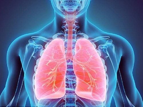
Pleural effusion, lobar pneumonia, ... are common respiratory syndromes and diseases. Recognizing the symptoms of some common respiratory syndromes and diseases will help patients plan timely examination and treatment, minimizing possible complications.
1. Pleural effusion
Pleural effusion is a condition in which fluid appears in the virtual spaces of the pleura.
1.1 Symptoms Depending on the amount of effusion, the symptoms of the disease will manifest in different degrees such as:
Less fluid: The patient has pain in the lung with effusion, can still lie on his back with his head low, however must lie on the side with the lung free of effusion to avoid pain. Moderate fluid: The patient has mild dyspnea and must lie on the side with the effusion lung. The patient has severe shortness of breath, shallow breathing, rapid breathing and cannot lie down, has to sit up to breathe. In addition, the disease also has other symptoms: fatigue, loss of appetite, high or low fever.
1.2 Diagnosis X-ray is the main method to confirm the diagnosis of pleural effusion. Depending on the amount of translation more or less, the image will have a large or small blurred area.
Little translation: Physical examination may be undetectable, but radiographs will show a blurred costophrenic angle. Mean translation: The X-ray image shows the Damoiseau curve. Massive effusion: One side of the chest has a blurred pleural effusion, showing widening of the intercostal space and poor mobility, the heart may be pushed to the right or left. Depending on the localized effusion in the interlobar, diaphragm or mediastinum, X-ray opacities will be shown corresponding to the site of effusion 3. In addition, pleural effusion can be performed in case of dyspnea due to the amount of media. It also helps to diagnose the cause of the effusion and then to have an appropriate treatment.

Tràn dịch màng phổi
2. Pneumothorax
Pneumothorax is one of the common respiratory diseases and syndromes, which can be caused by tuberculosis and is often a progressive complication of pulmonary tuberculosis. However, pneumothorax can also be seen in young people, including healthy people, the disease does not progress to pleural effusion, but often recurs, due to rupture of the pneumococcal cyst. Other known causes of pneumothorax are pertussis, a ruptured lung abscess that causes pneumothorax.
2.1 Symptoms Pneumothorax respiratory disease has the following symptoms:
Sudden sharp pain in the chest Shortness of breath Shock (due to mediastinal compression) 2.2 Diagnosis Pneumothorax can be detected by imaging Chest X-ray with the following images:
On the side of the lung, there is a hyperenhanced pneumothorax. The lungs contract and form a collapsed mass close to the hilum. The intercostal space is dilated, the diaphragm is poor or immobile, is pushed down, the mediastinum is pushed to the side of the uninjured lung.
3. Pulmonary consolidation syndrome
Pulmonary consolidation syndrome is a respiratory disease in which the lung parenchyma is spongy and has an increased density in a large or small area. When the lung parenchyma is inflamed, it will congestive damage to the alveoli, an area filled with secretions, leading to thickening of the lung parenchyma.
3.1 Etiology Non-tuberculosis pneumonia: Acute pneumococcal lobar pneumonia is the typical cause of pulmonary coagulation syndrome. The disease can occur at any age, especially those with weakened immune systems. The initial symptoms are usually sudden high fever, accompanied by chills, dry cough, chest pain, rapid pulse, slight shortness of breath, fatigue, loss of appetite, weight loss. Later, these symptoms get worse, with a lot of cough and sputum production. Subclinical diagnosis by blood test showed increased leukocytes, fresh microscopy, sputum culture can detect pneumococci. Chest X-ray shows opacities in one lobe or lobe of the lung.

Viêm phổi thùy là bệnh hô hấp thường gặp
Lung Abscess: Lung abscess is a respiratory disease in which the lung parenchyma becomes inflamed and festering due to some bacteria capable of causing pus. Chest X-ray showed opacities that were abscesses in the lungs. Pulmonary TB: Pulmonary tuberculosis can cause consolidation syndrome in one or more sites of the lung. The disease is likely to have a chronic progression with persistent febrile, debilitating symptoms. Performing a test of sputum in the throat can find the TB bacillus causing the disease. Atelectasis due to bronchial compression: Foreign bodies or blood clots can suddenly block large bronchi and cause atelectasis. Atelectasis respiratory disease has symptoms of shortness of breath, coughing up blood, and a collapsed thoracic cavity and poor mobility. Pulmonary infarction: Coagulation diseases such as mitral stenosis or after pelvic surgery, postpartum can block a branch of the pulmonary artery. Typical symptoms are shortness of breath, sudden chest pain, coughing up blood with purple-black color, shock, chest X-ray showing solidification. 3.2 Diagnosis Diagnosis of consolidation syndrome is mainly based on chest X-ray images with the following features:
Lesions are opacities occupying an area or scattered in the lung, with lobar pneumonia is an inflammatory image occupying a segment of the lung. lobe or one side of the lung. The opacity of the lung lesion is of uniform or irregular density with an indistinct or indistinct border. On the image, if the adjacent organs are seen to be pushed down, it helps to differentiate the pleural effusion.
4. Restrictive syndrome and obstructive syndrome
Restrictive syndrome and obstructive syndrome are common respiratory syndromes and disorders with manifestations of ventilation disorders and are determined by measuring ventilation function.
Restrictive syndrome is common in cases such as atelectasis, pulmonary fibrosis due to alveolar dilatation, obstruction, surgical removal of part of the lung.
Obstructive syndrome is common in cases such as bronchiolitis, asthma.
Additionally, mixed restrictive and obstructive syndromes can be seen in people with chronic obstructive pulmonary disease (COPD).
4.1 Upper Respiratory Obstruction Upper airway obstruction is a respiratory disease caused by causes such as:
Trauma: Jaw fracture, hypotonicity, hematoma, surgery, ... software damage oropharyngeal area, laryngeal trauma, esophageal foreign body. Pathology: Laryngeal edema or spasm, acute epiglottitis, acute laryngitis, laryngeal diphtheria, bilateral vocal cord paralysis, angioedema, coagulopathy. Allergies: Edema of the pharynx and trachea due to antibiotic allergy, bee stings. Tumor: Tumor of the larynx, purulent fossa in the oropharynx, obstructing the upper respiratory tract. Most respiratory syndromes and pathologies will have symptoms of shortness of breath. However, it is necessary to recognize the upper respiratory tract obstruction syndrome with other symptoms such as:
Slow or fast breathing, shallow breathing. In severe cases, you can choke or suffocate. Sweating, heavy can see consciousness disorder. The larynx or trachea hears a hissing sound. Auscultation of the lungs results in decreased alveolar murmurs on both sides of the lungs. accessory respiratory muscles are contracted. Late signs may include cyanosis of the lips and extremities. Upper respiratory tract obstruction can be diagnosed by radiography and laryngoscopy.
4.2 Lower respiratory obstruction Lower respiratory tract obstruction is a respiratory disease caused by causes such as:
The bronchi are compressed from the outside due to enlarged tracheobronchial nodes, pneumopericardium causing enlarged heart. The bronchi are compressed from the inside due to tumors (can be benign or malignant), foreign bodies, tuberculosis, diphtheria. Stagnation due to secretions such as coughing up blood, blood clots blocking the bronchi, bronchopneumonia.

Tắc nghẽn hô hấp dưới
Lower respiratory tract obstruction is also a respiratory disease, so the initial symptom is also shortness of breath, in addition, there are other signs such as:
During an asthma attack, it is often accompanied by cough and shortness of breath. Facial flushing, sweating, accessory respiratory muscles pulling like the wings of the nose. If auscultation of the lungs is completely obstructed, there is no alveolar murmur in the entire chest area; if the obstruction is incomplete, a crackling sound will be heard. Lower respiratory tract obstruction can be diagnosed by radiographs and shows atelectasis if the obstruction is complete. In case of incomplete blockage, air stasis will be seen because air can enter but cannot come out.
Respiratory diseases such as pneumonia, obstruction are mostly detected and diagnosed by X-ray. Some cases are caused by bacteria, viruses, ... need blood tests, fresh scans, sputum cultures to find bacilli.
Vinmec International General Hospital is a general hospital with the function of examining and treating common respiratory syndromes and diseases such as pneumonia, bronchitis, asthma, pleural effusion and many other diseases. other. At Vinmec, we have also performed endoscopic diagnosis and treatment with modern medical methods for respiratory diseases, not only bringing high efficiency but also minimizing complications of recurrent disease. The great success is because Vinmec is always fully equipped with modern facilities, examination and treatment procedures are carried out by a team of experienced and qualified doctors that bring optimal treatment results. advantage for customers.
If you have a need for consultation and examination at Vinmec Hospitals under the nationwide health system, please book an appointment on the website for service.
Please dial HOTLINE for more information or register for an appointment HERE. Download MyVinmec app to make appointments faster and to manage your bookings easily.


