This is an automatically translated article.
Posted by Master, Doctor Mai Vien Phuong - Department of Examination & Internal Medicine - Vinmec Central Park International General Hospital
Abbreviation
H. pylori-associated gastric cancer (HpPGC)
Stomach cancer not caused by H. pylori infection ( Stomach cancer not caused by H. pylori infection )
Helicobacter pylori ( H. pylori infection ) ) causes chronic inflammation, atrophy of the gastric mucosa and a high risk of developing gastric cancer. In recent years, awareness of H.pylori eradication treatment has increased in Vietnam. As H. pylori infection decreases, the incidence of gastric cancer arising from gastric mucosa that is not infected with H. pylori increases. The occurrence of gastric cancer arising in patients without H. pylori infection, although rarely reported, is a concern that needs to be addressed and should elucidate the clinical features of this disease. it.
1. How is the patient determined to be H. pylori-free?
The authors defined a patient as H. pylori-free using the following three criteria: (1) The patient was untreated for H. pylori, as determined by investigation of the medical record and performed present patient interviews; (2) no atrophy on endoscopy; and (3) The patient is negative for H. pylori after being tested at least twice by various diagnostic methods, including anti-H.Pylori -IgG serology, breath urease testing, rapid urease test, and microscopic examination.
2. Reports of gastric cancer not due to H. pylori infection
In Japan, a study of a total of 2462 patients with 3375 cases of early gastric cancer treated by endoscopic submucosal dissection was enrolled in the author's study since May. May 2000 to September 2019. Of these, 30 lesions in 30 patients were diagnosed as gastric cancer not due to H. pylori infection (Stomach cancer not due to H. pylori infection).
Results of the study:
The frequency of stomach cancer not due to H. pylori infection Stomach cancer not due to H. pylori infection was 1.2% (30/2462) for the patients in the study. The study included 19 men and 11 women with a mean age of 59 years. The location of gastric lesions is divided into three parts; Upper 1/3 (U), middle 1/3 (M), lower 1/3 (L). Of the 30 lesions, 15 were U, 1 were M, and 14 were L. Morphologically, 17 were convex and flat (0-I, 0-IIa, 0-IIa + IIc), and 13 flat lesions and depressive type (0-IIb, 0-IIc). The mean diameter of the tumor was 8mm (range 2-98mm). Histological analysis showed that 22 lesions (73.3%) were differentiated type. Stomach cancer lesions not due to H. pylori infection Stomach cancer not due to H. pylori infection was classified as basal adenocarcinoma (7 cases), well differentiated carcinoma (8 cases) cases), intestinal phenotype carcinoma (7 cases) and ring cell carcinoma alone (8 cases). Among 30 patients with gastric cancer not due to H. pylori Stomach cancer not due to H. pylori infection, 24 lesions (80%) were confined to the mucosa; in which, the remaining 6 lesions showed submucosal invasion.
The authors elucidated the clinical pathologic features of gastric cancer not due to H. pylori infection, they found that the cancer types are not only differentiated but also undifferentiated. . In addition, intestinal phenotype tumors were also observed and may be an important feature.

Người mắc ung thư dạ dày cần được thăm khám và phát hiện sớm giúp điêu trị thành công
3. H.pylori – a risk factor for stomach cancer
Most stomach cancers are associated with infection with the bacterium Helicobacter pylori (H. pylori) during their development, and in 1994, H. pylori was certified as a “determined carcinogen.” for gastric cancer development. H. pylori infection leads to inflammation, atrophy of the gastric mucosa and intestinal metaplasia; When H. pylori infection becomes chronic, there is a high risk of stomach cancer.4. Stomach cancer not caused by H. pylori infection
In recent years, awareness of eradication treatment has been enhanced in Japan, thereby reducing the prevalence of H. pylori infection, especially among young people due to improved sanitation and expansion of only eradication. As H. pylori infection decreases, the incidence of gastric cancer arising from gastric mucosa not infected with H. pylori increases. Currently, however, gastric cancer not caused by H. pylori infection is very rare compared to H. pylori-associated gastric cancer (HpPGC). This definition is still not well established and its frequency is reported to vary from 0.4 to 5.4%.
Previous studies have reported that undifferentiated H. pylori gastric cancer was observed more frequently than undifferentiated type . However, in recent years, differentiated gastric cancers such as adenocarcinoma, even including gastric cancer not caused by H. pylori infection have been reported. Very few studies have investigated gastric cancer that is not caused by H. pylori infection and its clinical features are not well documented. Therefore, it is necessary to elucidate the characteristics of gastric cancer not due to early H. pylori infection.
5. Characteristics of clinical features of gastric cancer not caused by H. pylori infection
A total of 2462 consecutive patients with 3370 c gastric cancers (3132 early gastric carcinoma lesions and 238 adenomas) were enrolled. A total of 30 lesion types 30 patients (1.2%) were classified as gastric cancer not due to H. pylori infection. The study included 19 men and 11 women with a mean age of 59 years. Of the 30 lesions, 15 were U, one M, and 14 L. Morphologically, 17 were convex and flat (0-I, 0-IIa, 0-IIa + IIc), and 13 flat and concave lesions. type (0-IIb, 0-IIc). Tumor diameters ranged from 2 mm to 98 mm, with a mean diameter of 8 mm. Histopathologically, 22 lesions (73.3%) were identified as differentiated and 8 lesions (26.7%) were undifferentiated. All tested undifferentiated lesions were squamous cell carcinomas. Tumor invasion in 24 lesions (80%) was limited to the mucosa, while the remaining 6 lesions showed submucosal invasion. One of the lesions invaded the submucosa to a depth of 500 μm (SM2). Outcomes for patients with gastric cancer not due to H. pylori infection were positive, all were successfully resected, with no tumor margins.
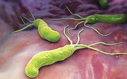
H. pylori có thể gây ung thư dạ dày ở một số người bệnh
6. Frequency of gastric cancer not due to H. pylori infection
H. pylori infection causes chronic inflammation and atrophy of the gastric mucosa and often leads to gastric cancer. Since 1994, H. pylori has been recognized as a “certain carcinogen” that contributes to the development of gastric cancer. A prospective study reported that H. pylori eradication therapy prevented two-thirds of cases of gastric cancer.
Therefore, since 2010, the Japanese insurance system has allowed patients who have undergone laparoscopic tumor resection to receive H.pylori eradication therapy, and in current H. pylori eradication therapy. of Japan is the insurance treatment for all patients infected with H. pylori . Improved sanitation has significantly reduced the incidence of new H. pylori infections and H. pylori infection rates in young adults are reported to decrease annually.
The frequency of gastric cancer not due to H. pylori infection is low; however, this number is expected to increase and the frequency of Stomach Cancer not due to H. pylori infection may increase accordingly. Currently, gastric cancer that is not caused by H. pylori infection is still rarely reported to date, and the frequency varies considerably from 0.66% to 14% of gastric cancer cases. The difference in this range may be due to differences in the definition of H. pylori-free status in previous reports.
7. Role of NBI in gastric cancer diagnosis
In recent years, the diagnosis of NBI has become indispensable in the diagnosis of gastric cancer. In Japan, Yao's VS (microchip architecture and microsurface structure) classification system is widely cited. Furthermore, Yokoyama et al's classification is useful as it can aid in early gastric cancer identification in NBI imaging. However, in these patients with gastric cancer not due to H. pylori infection, there are many cases that cannot be characterized by the conventional NBI classification of gastric cancer. For example, in basal adenocarcinoma, the demarcation line is not clear, because the tumor is covered with non-neoplastic epithelium, the surface microstructural pattern is frequent NBI findings and typical of such differentiated adenocarcinoma could not be observed.
In addition, pure ring cell carcinoma does not exhibit the typical helical pattern as mentioned above. These cases are classified according to the NBI classification system. Therefore, gastric cancer not caused by H. pylori infection may not fit the conventional NBI classification system.
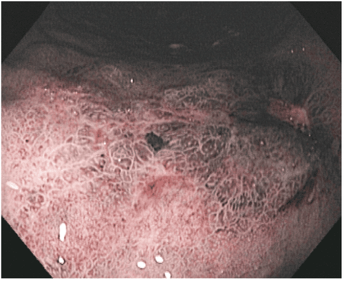
Nội soi bằng phương pháp NBI giúp bác sĩ chẩn đoán sớm ung thư dạ dày
Conclusion
Recognizing the different clinical features of gastric cancer not due to H. pylori infection is very useful for future clinical diagnosis. Because gastric cancer not due to H. pylori infection is a rare occurrence, the authors are considering a multicenter clinical trial to collect cases to elucidate the clinical features in more detail. of gastric cancer not due to H. pylori infection. Multicentre observational trials are the best method for future research.
Currently, Vinmec International General Hospital is a prestigious address trusted by many patients in performing diagnostic techniques for digestive diseases, diseases that cause chronic diarrhea, Crohn's disease, gastric mucosa Esophageal ectopic... Along with that, at Vinmec Hospital, screening for stomach cancer and gastric polyps is done through gastroscopy with Olympus CV 190 endoscope, with NBI (Narrow) function. Banding Imaging - endoscopy with narrow light frequency) results in clearer images of mucosal pathology than conventional endoscopy, detecting ulcerative colitis lesions, digestive cancer lesions Early stage...
Vinmec Hospital with modern facilities and equipment and a team of experienced specialists who are always dedicated in medical examination and treatment, customers can be assured of internal services. gastroscopy and esophagoscopy at Vinmec International General Hospital.
Please dial HOTLINE for more information or register for an appointment HERE. Download MyVinmec app to make appointments faster and to manage your bookings easily.
ReferencesUemura N, Okamoto S, Yamamoto S, Matsumura N, Yamaguchi S, Yamakido M, Taniyama K, Sasaki N, Schlemper RJ. Helicobacter pylori infection and the development of gastric cancer. N Engl J Med. 2001;345:784-789. [PubMed] [DOI] Schistosomes, liver flukes and Helicobacter pylori. IARC Working Group on the Evaluation of Carcinogenic Risks to Humans. Lyon, 7-14 June 1994. IARC Monogr Eval Carcinog Risks Hum 1994; 61: 1-241. [PubMed] Parsonnet J, Vandersteen D, Goates J, Sibley RK, Pritikin J, Chang Y. Helicobacter pylori infection in intestinal- and diffuse-type gastric adenocarcinomas. J Natl Cancer Inst. 1991;83:640-643. [PubMed] [DOI] Yamamoto Y, Fujisaki J, Omae M, Hirasawa T, Igarashi M. Helicobacter pylori-negative gastric cancer: characteristics and endoscopic findings. Dig Endosc. 2015;27:551-561. [PubMed] [DOI] Chiko Sato, Kingo Hirasawa et al., Clinicopathological features of early gastric cancers arising in Helicobacter pylori uninfected patients, World J Gastroenterol. May 28, 2020; 26(20): 2618-2631