This is an automatically translated article.
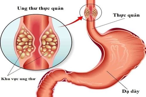
Post by Master, Doctor Mai Vien Phuong - Gastrointestinal Endoscopy - Department of Medical Examination & Internal Medicine - Vinmec Central Park International General Hospital.Colorectal cancer is a major health problem, representing the second most commonly diagnosed cancer in women and the third in men. Today, colonoscopy is the most used tool for early detection of colorectal tumors as well as for resection of precancerous lesions to prevent late-stage and advanced cancers.
In fact, it is well known that over 95% of colorectal cancers arise from adenomas (tumours of benign neoplastic epithelium with variable malignancy) and the purpose of endoscopic follow-up colon is to interrupt the “sequence of adenocarcinoma”.
1. Endoscopists are increasingly interested in pedunculated polypoid lesions In recent years, there has been growing interest in non-polyposis colorectal tumors (pedicleless polyps) and in particular polyps. lateral spread (LST). LST is a large flat neoplasm that tends to grow along the surface of the intestine. By definition, LST indicates a diameter in excess of 10 mm. LSTs and large sessile polyps have a higher frequency of high-grade dysplasia (HGD) and are more locally invasive than nodular lesions of the same size and are often a technical challenge for the endoscopist. both diagnostic and resection. That is why the term “advanced mucosal neoplasm” (sessile neoplasm) has been proposed recently for these two types of lesions.
2. Research and classification of colorectal tumors
The morphology of colonic lesions depends on the proliferative direction of the tumor. Then, two major macrotypes can be recognized: Superficial (type 0) and advanced (type 1-5) cancers.
According to the Paris classification, superficial lesions (type 0) are distinguished in: Polyp type [2.5 mm higher than the pedunculated mucosa (0-1p), pedunculate (0-) 1s) or mixed (0-1sp)]; polyps are flat [slightly less than 2.5 mm (0-IIa), flat (0-IIb) or slightly concave (0-IIc)] and mixed types.
Paris classification of superficial colorectal polyps :
Polyp type: Stemless (0-1p), Stemless (0-1s), Mixed (0-1sp) Flat: Slightly raised (0) -IIa), Flat (0-IIb), Slightly Concave (0-IIc) Mixed Types: High and Concave (0-IIa + IIc), Concave and High (0-IIc + IIa) Threshold 2.5 mm , corresponding to the height of the biopsy forceps when closed is quite arbitrary and not really reliable because many flat lesions are not uniform over their entire surface.

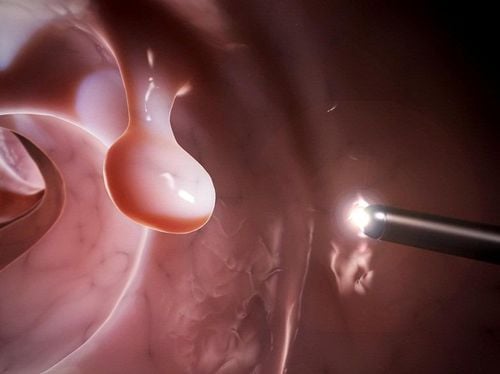
Polyp dạng phẳng LST được phát hiện qua nội soi
Stemless polyps (flat or concave) can be found throughout the colon, unlike pedunculated polyps that are more common on the left side.
It is not always easy to grasp the distinction between the different subtypes, so the local endoscopist and pathologist play an important role in the diagnostic algorithm.
As noted above, flat colorectal tumors that are equal to or larger than 10mm in size are called LSTs.
4. Classification of lateral polyps LSTs are divided into granular (LST-G) and non-granular (LST-NG) based on their detailed endoscopic images during endoscopic staining with spray carmine indigo dye. The LST-G type consists of agglomerations of nodules forming a flat mass over a wide surface (hence the term "granular") while this property is lacking in the latter group (LST-NG).
Stemless polyps have a higher risk of local invasion than pedunculated polyps, regardless of size. Although the larger the polypoid lesion, the higher the risk of invasive submucosal disease, not all sessile polyps show a strong correlation between size and local invasion.

Các dạng polyp lan sang bên
LST-Gs with a uniform surface have a low risk of invasive submucosal disease (<2%), regardless of their size, while LST-Gs with mixed size nodules are at risk. The risk of submucosal invasion was higher (7.1% for lesions < 20 mm and 38% for those > 30 mm). The risk of submucosal invasion for LST-NGs was even higher, especially in polyps with thinner centers (LST-NGs with pseudoconcave) 12.5% for size <20 mm and 83.3% for diameters > 30 mm.
Concave lesions are rare (1%-6% of all pedunculated polyps) but have the highest risk of submucosal invasion at 27%-35.9%.

Các dạng phụ của dạng polyp không cuống theo phân loại Paris. Các polyp dạng phẳng (polyp không cuống ) ≥ 1 cm được gọi là các khối u lan sang bên (LST).
5. Endoscopic diagnosis The discovery of mucosal surface lesions in asymptomatic patients undergoing total colonoscopy is a frequent event, ranging from 10% to 60%.
In a Japanese series, the incidence of sessile colon polyps was 42% (10,948 out of 25,862 identified superficial cancerous lesions). This percentage is lower, though still significant, in another series of Japanese studies (27%: 2711/12811). In the United States and Western countries, the prevalence of sessile polyps varies widely, from 9.35% to 31.4%.
Stemless polyps can often be missed by inexperienced endoscopists, so careful training and accurate assessment of all suspicious mucosal areas is required.
Coloroscopy or narrow band imaging (NBI) should be considered to estimate the risk of sessile polyps of submucosal invasion. The NBI technique, which uses a light filter to narrow the endoscope's light bandwidth to selectively evaluate the area of interest, can better recognize and define vascular and pituitary patterns, both The second is a sign of malignancy. For example, irregular, sparse vascularization and loss of epithelial crests are associated with progressive lesions.
Kudo et al have extensively described the microarchitecture of pits, epithelial crests or ridges (also known as “pits models”): Three main types of pit patterns are described: Nonplastic (I and II); Neoplastic adenomas (III and IV); Cancerous tumor (V).
Surface polyps are classified into the following types:
Non-neoplastic: I - Normal mucosa, II-Regularly enlarged pits Neoplastic, adenoma: IIIL - Long banded pits , IIIS - Narrow round and irregular pits, IV- Branched pits. Neoplasm, cancer: Vi- Irregular surface, VN-Amorphous surface. With regard to vascular circulation, irregular multi-branch microvasculature interspersed with avascular regions is a predictor of a higher risk of submucosal invasive disease.
Types of vascular patterns on the surface of the colonic mucosa:
Not neoplastic: Normally, capillaries are clearly delineated around the opening; Faint, poor visibility of capillaries around enlarged fossa. Neoplasm, adenoma: A network of blood vessels arranged in a regular grid; Dense - enlarged blood vessels of regular size. Neoplasm, cancer: Infrequently with large blood vessels of irregular diameter and direction of division; Sparsely poorly distributed irregular circuits with divergent directions. Even when no single feature is absolutely specific for submucosal invasion, the presence of more than 1 high-risk feature correlates with more invasive lesions.
LST-Gs account for 60%-80% of cases compared with 20%-40% of LST-NGs, while concave sessile polyps (those with a higher risk of submucosal invasion) account for 1% to 6% of all superficial colorectal lesions. Most studies reported the majority of sessile polyps in the right colon (55.7%-80%), different from the two Chinese series, where 65%-75% of non-polyposis lesions were present. The stalk is located in the left colon.
6. Conclusion Narrow polyps (NPTs) are distinguished as slightly elevated (0-IIa, less than 2.5 mm elevated), flat (0-IIb) or slightly concave (0-IIc). Stemless polyps are usually flat or slightly elevated, whereas concave lesions suggest an increased risk of submucosal invasion (SMI). Coloroscopy or narrow band imaging should be considered to estimate the risk of pedunculated polyps of submucosal invasion and characterize the pituitary and vascular patterns to predict the risk of invasion. . Endoscopic mucosal resection remains first-line therapy for sessile polyps, while surgery or endoscopic submucosal dissection should be considered for larger tumors presenting with submucosal invasion. desert.
Please dial HOTLINE for more information or register for an appointment HERE. Download MyVinmec app to make appointments faster and to manage your bookings easily.