This is an automatically translated article.
Articles written by MSc, BS. Dang Manh Cuong, Department of Diagnostic Imaging - Vinmec Central Park International General Hospital
Thoracic resonance imaging is a non-invasive, non-radiative imaging technique that provides clear, detailed images of the heart and vascular structure that other imaging methods cannot. can do.
1. What is magnetic resonance imaging (MRI) of the chest?
Magnetic resonance imaging (MRI) is a noninvasive examination used to diagnose medical conditions. An MRI uses a strong magnetic field, radio waves, and a computer to create detailed images of structures inside the body. An MRI does not use radiation (X-rays). Detailed MRI images allow doctors to evaluate normal and pathological structures inside the body. Images can be played back on a computer monitor. They can also be sent in print or copied to a CD or uploaded to the digital cloud. Thoracic MRI provides detailed images of structures within the thorax, including the chest wall, pleura, mediastinum, heart, and mediastinal vessels at almost any angle. MRI also provides film-like images of the cardiac system and blood vessels, which is important for evaluating the integrity and function of these structures (heart, heart valves, great vessels, etc.).

Chụp MRI là kỹ thuật chẩn đoán hình ảnh an toàn, chính xác
2. What is the chest MRI for?
MR imaging of the chest is done to:
Evaluate abnormal masses (including lung cancer or other cancers) that cannot be adequately assessed with other imaging modalities (usually CT) or cases that can only be assessed by MRI. Determine tumor size, extent of spread, and extent of invasion to adjacent structures. Assess the anatomy and function of the heart and other heart structures (heart valves, etc.). Assess myocardial perfusion (blood flow to the heart) and myocardial infarction (scarring in the heart muscle due to previous blockage of blood vessels supplying the heart). Determination of flow kinetics in blood vessels and chambers of the heart. Shows lymph nodes and blood vessels, including vascular and lymphatic malformations of the thorax. Evaluation of disorders of the thoracic system (vertebrae, ribs, and sternum) and soft tissue of the chest wall (muscle and fat). Evaluation of pericardial disease. Characterize mediastinal or pleural lesions seen with other imaging modalities, such as chest X-ray or CT. A special type of MRI called magnetic resonance angiography (MRA) is useful for evaluating the blood vessels in the chest (both arteries and veins). MRA can also clearly show aneurysms or dissection of the blood vessel wall.

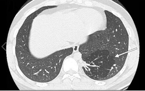
Chụp MRI lồng ngực giúp tầm soát bệnh ung thư phổi
3. Does the patient need to prepare anything? How to prepare?
You will need to wear a hospital gown or may be able to wear your own clothes if it is loose and does not have metal fasteners.
Your doctor will give you specific instructions on what to eat and drink before the MRI scan.
If contrast injection is indicated, you will be asked if you have asthma or are allergic to iodinated contrast agents, drugs, foods or the environment? Because in contrast medicine, there is a contrast agent called gadolinium. Gadolinium may be used in patients with an iodine contrast allergy. Patients are less likely to be allergic to gadolinium contrast than to iodinated contrast. However, even if the patient is known to be allergic to gadolinium, it can be used after notifying the doctor for consideration.
If you have any serious health problems or have had recent surgery, tell the technician or radiologist. Certain conditions, such as severe kidney disease, may require the use of special gadolinium contrast agents that are considered safe for patients with kidney disease. You may need kidney function tests to determine if your kidneys are working properly.
If you are a woman, you should always tell your doctor and technician if you are pregnant. MRI has been used since the 1980s but there have been no reports of any adverse effects on pregnant women or their unborn babies. However, the baby will be in a strong magnetic field, so pregnant women in the first 3 months should not have an MRI unless the benefits of the scan clearly outweigh the potential risks. Pregnant women should not have gadolinium contrast injections unless absolutely necessary.
If you have claustrophobia (fear of confined spaces) or are very anxious, you can ask your doctor to prescribe a mild sedative before your exam.
Infants and young children often require sedation or anesthesia to complete an MRI without mobility. This depends on the child's age, intellectual development and type of shooting. A specialist in pediatric sedation or anesthesiology will be present during the scan for your child's safety. You will be told how to prepare for your child.
Leave all jewelry and other accessories at home or remove them before the MRI. Metal and electronic items can interfere with the magnetic field of the MRI machine and they are not allowed in the imaging room. They can cause burns or become harmful in the MRI machine room. These include:
Jewelry, watches, credit cards, and hearing aids all of which can be damaged Pins, hairpins, metal zippers and similar metal objects, can cause disfiguration. distortion of the MRI image. Removable dentures Pens, pocket knives and eyeglasses Body piercings Cell phones, electronic watches and tracking devices.
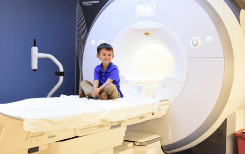
Trẻ em khi chụp MRI cần sử dụng thuốc an thần
In most cases, an MRI is safe for patients with metal implants, with some exceptions. People with the following implants may not be screened and should not enter the MRI area without first being evaluated for safety:
Certain cochlear implants Certain types of clips are used for aneurysms brain Some types of metal coils are placed in blood vessels Some older defibrillators and pacemakers Tell the technician if you have medical or electronic equipment in your body. These devices may interfere with the shooting process or pose a risk. Many implantable devices will have a brochure explaining the MRI risks for that particular device. If you have the pamphlet, bring it to your doctor before the scan is done. MRI cannot be performed without confirmation and documentation of implant type and compatibility with MRI.
If there is any question, X-ray can detect and identify any metal object. Metal objects used in orthopedic surgery usually pose no risks in an MRI. However, a recently placed artificial joint may require the use of a different imaging technique.
Tell the technician or radiologist about any shrapnel, bullets, or other metal that may be in your body. Foreign objects near and in particular in the eye are important because they can move or heat up during the scan and cause blindness. The dye (tattoo ink) used in tattoos may contain iron and can heat up during the MRI scan and cause burns. This is rare.
Fillings, braces, eyeshadows and other cosmetics are generally unaffected by magnetic fields. However, they can distort images of the face or brain area. Tell the radiologist about them.
Anyone accompanying the patient into the MRI room must also be examined for metal objects and implanted devices.

Đeo niềng răng khi chụp MRI có thể ảnh hưởng đến kết quả chụp
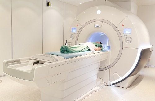
4. How is the process done?
Thoracic MRI can be performed on both inpatients and outpatients. You will be placed on a movable table. Straps and splints may be used to help you lie still and keep your position. Place devices containing coils capable of sending and receiving radio waves (coils) around the area of the body being scanned. You will be placed inside an MRI machine. The technician will perform the scan while sitting in the control room outside the MRI machine room. An MRI scan usually involves several runs (pulses), each of which can last several minutes. If magnetic contrast medicine is used, your doctor, nurse, or technician will place a line into a vein in your hand or arm to inject contrast. Once the scan is complete, you can lie down for a while while the radiographer checks the quality of the images in case you need to take another or more pictures. An intravenous line in your arm will be removed after the scan is over. The whole shooting process will be completed in about 20-30 minutes.

Thời gian chụp cộng hưởng từ diễn ra nhanh chóng
5. What was your experience during and after the MRI scan?
Most MRI scans are painless. However, there are some patients who are uncomfortable for the following reasons:
Feeling claustrophobic in the MRI scanner (clautrophobic syndrome). Therefore, some patients with anxiety need sedation. Feeling noisy when the MRI machine pulses The area you lie on may feel a little hot, but that is normal. If it bothers you, tell your doctor or technician. It is important that you remain completely still during the shoot. You'll know when the image is being recorded because you'll hear and feel the loud knocking as the coils that generate the radio frequency pulses are activated. Some centers provide earplugs, while others use music headphones to reduce the intensity of the sound produced by the MRI machine. You should be able to relax between pulse bursts, but still have to stay in your position without moving as much as possible.
You will usually be alone in the MRI room. However, the technician can still see, hear and talk to you at all times using the two-way intercom. If you are concerned about needing a loved one by your side, you can request that as long as they are also checked for safety before entering the MRI room.
Children will be fitted with properly sized earplugs or headphones during the search. Music can be played through headphones to help you forget about long periods of shooting.
If you have an appointment for an intravenous contrast scan, it is normal to feel a hot flush for a minute or two after the injection.
The IV can cause you some discomfort when it is inserted and removed, you may experience some bruising. There is also very little chance of irritating your skin at the site of the IV placement.
If you are not under anesthesia, there is no need for follow-up time after the scan. You can resume your normal activities and diet right after the scan.
Some patients experience side effects from the contrast agent, including nausea and focal pain. Very rarely, the patient is allergic to the contrast agent and develops urticaria, itchy eyes, or other allergic reactions. If you experience allergy symptoms, a radiologist or other doctor will be ready to take action right away.
6. What are the benefits and risks of this approach?
6.1 Benefits
MRI is a non-invasive, non-radiative imaging technique. MR images of the heart and vascular structures are often clearer and more detailed than with other imaging methods. This detail makes MRI an invaluable tool in the early diagnosis and evaluation of cardiovascular conditions. MRI has proven valuable in the diagnosis of a wide range of conditions, including cancer, heart and blood vessel disease, heart valve abnormalities, and other bone and soft tissue abnormalities of the chest. MRI is also useful for staging the tumor. An MRI can help doctors evaluate both the structure of an organ and how it is functioning. The contrast agent gadolinium used in MRI scans is less likely to cause allergic reactions than the iodinated contrast agent used for X-rays and CT scans. Chest MRI is generally more informative than other imaging methods, with the exception of lung parenchymal abnormalities, where chest CT is the preferred imaging test. MR imaging can assess blood flow without the risk of side effects of conventional angiography.
Chụp cộng hưởng từ rất ít khi gây ra các tác dụng phụ
6.2 Danger
MRI scans are virtually risk-free for the normal patient as long as safety instructions are followed as strong magnetic fields are not harmful. However, it can cause implanted medical devices to malfunction or cause image distortion. Renal system fibrosis is a recognized, but rare, complication associated with injection of Gadolinium contrast agent. It usually occurs in patients with severe kidney disease. Your doctor will evaluate your kidney function before injecting the contrast agent. There is very little risk of an allergic reaction if a contrast agent is used. Such reactions are usually mild and controlled with medication. If an allergic reaction occurs, a doctor will be available to assist immediately. The manufacturers of intravenous contrast agents say that mothers should not breastfeed for 24-48 hours after the contrast injection. However, the most recent ACR Manual of Contrast Contrast reports that studies have shown that the amount of contrast absorbed by infants during breastfeeding is extremely low.
7. What are the limitations of chest MRI?
Getting a clear shot depends a lot on your ability to lie still during the shoot and follow the instructions to hold your breath while recording the image. Therefore, if you are worried, confused or in severe pain, it is difficult for you to lie still during the scan, so the image quality will be low and affect the doctor's reading of the film. A person who is too large may not fit on some types of MRI machines. Implants and other metal objects can cause photographic artifacts. Patient movements can blur the image. Very irregular heart rhythms can also affect image quality. This is because some imaging techniques are based on the electrical activity of the heart. MRI is generally not recommended for patients with severe trauma. However, the decision to have imaging should be based on clinical judgment. This is because immobilizers and life support equipment may not be brought into the imaging room or, if brought in, may distort the MRI image. Therefore, they must be placed away from the area to be photographed. However, some trauma patients may need an MRI if necessary. Although there is no evidence that MRI harms the fetus, women in the first trimester of pregnancy should not have an MRI unless: necessary loss. MRI is often more expensive and takes longer to capture than other imaging methods. A chest MRI takes longer than an X-ray or CT scan. Because the MRI takes a long time, many children and infants need sedation to stay still during the scan.

Hệ thống máy chụp cộng hưởng từ MRI 3.0 Tesla hiện đại tại Bệnh viện Vinmec Hải Phòng
Vinmec International General Hospital put into use the magnetic resonance imaging machine 3.0 Tesla Silent technology. Magnetic resonance imaging machine 3.0 Tesla with Silent technology of GE Healthcare (USA).
Silent technology is especially beneficial for patients who are children, the elderly, weak health patients and patients undergoing surgery. Limiting noise, creating comfort and reducing stress for customers during the shooting process, helping to capture better quality images and shorten the shooting time. Magnetic resonance imaging technology is the technology applied in the most popular and safest imaging method today because of its accuracy, non-invasiveness and non-X-ray. Customers please make an appointment at the website or contact Vinmec Health System nationwide for service.
Please dial HOTLINE for more information or register for an appointment HERE. Download MyVinmec app to make appointments faster and to manage your bookings easily.
Reference source: radiologyinfo.org