This is an automatically translated article.
The article was professionally consulted by Specialist Doctor I Tran Cong Trinh - Radiologist - Radiology Department - Vinmec Central Park International General Hospital. The doctor has many years of experience in the field of diagnostic imaging.According to statistics of many countries, cardiovascular diseases, especially congenital heart disease, are the cause of high mortality. In recent years, many advances in congenital heart diagnosis such as cardiac magnetic resonance imaging have helped to detect the disease early, thereby providing a better chance of survival and prolonging life for patients.
1. What is Magnetic Resonance Imaging?
Magnetic resonance imaging (MRI) is a method that uses a strong magnetic field, radio waves and a computer to image parts of the body, helping to diagnose many different diseases. Cardiac magnetic resonance imaging is an imaging technique widely used in the diagnosis of patients with or suspected cardiac disease.The advantages of cardiovascular MRI are high soft tissue contrast, high spatial resolution, multiple cross-sections, no radiation, and non-invasiveness. The whole scan doesn't have the same side effects as an X-ray or CT scan, but it still has the ability to detect abnormalities behind the layers of bone - something other imaging methods can't do. At the same time, this method also gives faster and more accurate images than X-rays in the diagnosis of cardiovascular diseases. At present, cardiovascular MRI technique has been widely used in coronary artery disease, heart valve, myocardium, congenital heart, heart failure,...
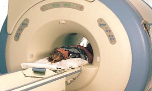
Chụp cộng hưởng từ tim là kỹ thuật không xâm lấn, an toàn cho người bệnh
2. Cardiac magnetic resonance imaging procedure
Cardiac magnetic resonance plays an important role in the diagnosis and monitoring of cardiovascular diseases.2.1 Indications and contraindications
IndicationCongenital heart disease: Evaluation of anatomical abnormalities, shunt,... Common congenital heart diseases include atrial septal defect, ductus arteriosus, tetralogy of Fallot, outflow obstruction right ventricle, coarctation of the aorta,... Cardiomyopathy: Myocardium dilated, myocardium hypertrophy, myocardium untrafted, myocarditis, iron deposition in myocardium,... Coronary artery disease : Evaluation of left ventricular function and movement, assessment of myocardial survival in myocardial infarction; Primary and secondary cardiac tumors: Provides tumor properties, helps to confirm diagnosis, evaluate the invasion of the tumor; Other pathologies: Heart valve disease, pericardial disease, metabolic cardiomyopathy.
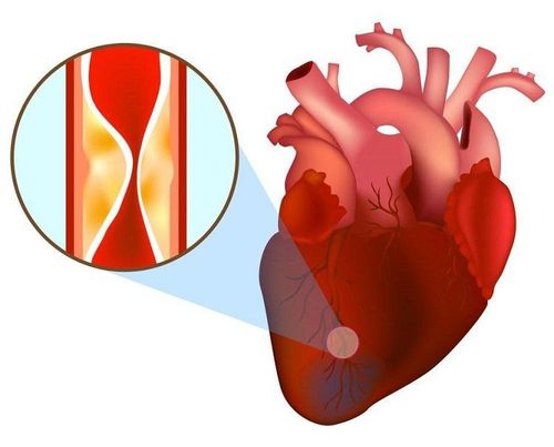
Bệnh nhân nhồi máu cơ tim được chỉ định thực hiện chụp cộng hưởng từ tim
Patients with implanted metal devices (pacemakers, defibrillators) need to be removed before the scan because the high magnetic field of the MRI machine can harm these devices; People with a fear of being in closed spaces; Person with a metal foreign body in the skull; Pregnant women in the first 3 months.
2.2 Preparation before implementation
Personnel: Specialist doctors, radiology technicians and nurses; Technical facilities: Magnetic resonance imaging machine, film, film printer and image storage system; Medical supplies: IV needles, syringes, distilled water (or physiological saline), cotton, gauze, gloves, sterile adhesive bandages, medicine boxes, emergency equipment for contrast drug accidents; Drugs: Magnetic contrast agents, sedatives and skin antiseptics; Patient: Thorough explanation of the procedure, no fasting, checked for necessary contraindications, instructions on changing clothes from the MRI room, removal of contraindicated items, receipt request magnetic resonance imaging or complete medical records if needed.
Bệnh nhân có thể được chỉ định tiêm thuốc an thần trước khi chụp MRI tim
2.3 Implementation process
Patient position: Lying supine on the magnetic resonance imaging table; selection and positioning of signal coils; move the table into the area of the MRI machine compartment, locate the imaging area; Location capture; Depending on the pathology and clinical requirements for cardiac magnetic resonance imaging according to appropriate procedures; Take pulse sequences to evaluate the morphology and function of the heart; Magnetic contrast injection to assess myocardial perfusion, evaluate drug kinetics through the cardiac chambers; Inject the drug in the late phase after 10 minutes, assess the survival of the myocardium; Electro-optical technicians process images, print films and transfer images and data to doctors; The doctor analyzes the obtained MRI images.2.4 Evaluation of results
The obtained images clearly show the anatomical structures of the coronary arteries and the heart; Accurate assessment of heart function.
Hình ảnh MRI tim được xử lý qua phần mềm và in phim
2.5 Complications and how to deal with them
The patient is scared and agitated: The doctor should encourage and comfort the patient; Patients are too worried, scared: The doctor can give the patient a sedative under the supervision of an anesthesiologist; Complications related to contrast agents: Although not toxic to the body, it can cause mild allergies with manifestations such as rash, numbness in limbs, nausea, headaches, ... or can also cause severe or severe reaction such as anaphylaxis. Depending on each specific case to have the appropriate treatment according to the standard protocol. Cardiac magnetic resonance imaging is the most modern method of diagnosis and monitoring of cardiovascular diseases today. Thanks to this technique, doctors can predict and provide effective treatment for congenital heart patients, thereby stopping the progression of the disease, improving the chances of curing the disease or prolonging the life of the patient. sick.Currently, Vinmec International General Hospital is equipped with a 640-slice CT scanner TSX - 301C manufactured by Toshiba capable of supporting diagnosis on an area up to 16 cm wide, fast speed allows one rotation of the image. The whole heart can be detected, helping to optimally diagnose coronary, vascular and systemic arteries. In particular, this machine can reduce up to 90% of radiation dose, so it can be taken for pregnant women when indicated.
Please dial HOTLINE for more information or register for an appointment HERE. Download MyVinmec app to make appointments faster and to manage your bookings easily.


