This is an automatically translated article.
The article was professionally consulted by Specialist Doctor I Nguyen Thanh Hai - Radiologist - Department of Diagnostic Imaging and Nuclear Medicine - Vinmec Times City International General Hospital. Doctor Hai has more than 20 years of experience in the field of diagnostic imaging, especially in the field of multi-slice computed tomography, magnetic resonance.Disc herniation is a disease caused by the patient's mucus coming out of the disc capsule, then compressing some other organs and causing pain to the patient, so X-rays cannot detect the disease at a mild stage. When this part of the nucleus pulposus has spilled out a lot, compressing the nerve roots, encroaching on the spinal cord, and deforming the vertebrae, X-rays can help the doctor diagnose the disease.
1. What is a herniated disc?
Herniated disc is a disease that is not directly life-threatening, but it causes serious effects on the patient's mobility as well as health.The main cause of disc herniation is because the disc's mucus nucleus is displaced from its original position, leading to compression of the nerves and prolonged pain for the patient. In addition, some other risk factors for disc herniation are due to:
Due to age: Because of the aging process, the discs and spine are dehydrated, sclerosed, and very easily damaged. injury leading to disc herniation. Due to injury in the back area, labor, movement and overwork, wrong posture, leading to damaged discs and spine Congenital diseases in the spine area or genetics. Obesity: The greater the body weight, the more pressure on the spinal discs, especially in the lumbar region, causes herniated discs. Herniated discs, if not treated early, will leave serious complications such as: Compressing nerve roots, narrowing the spinal cavity so that the patient can spend; lumbar nerve roots are compressed, causing uncontrolled defecation; Long-term sedentary causes muscle weakness, atrophy, reduced mobility; Urinary sphincter dysfunction leads to urinary retention, bedwetting,...

2. Can X-ray detect disc herniation?
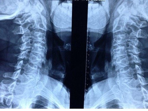
X-ray is a method of taking pictures from the outside of the body and still seeing changes inside of bones, joints and other organs. Because the X-ray machine emits high beams of X-rays, these X-rays penetrate soft tissues and body fluids to create images. From these images, the doctor will diagnose the disease. However, dense tissues such as bone will block X-rays a lot, if the tissue is highly dense, X-rays will be less penetrating.So can X-ray detect disc herniation? This is a disease that occurs when the nucleus pulposus comes out of the disc capsule, then compresses some other organs and causes pain in the patient. Therefore, X-rays cannot detect the disease at a mild stage because the spinal structures are less affected.
What is the use of X-ray in herniated disc disease? Accordingly, the X-ray can only:
Diagnose the disease when this part of the nucleus pulposus has overflowed, compressing the nerve roots, has vertebrae deformity, and narrows the intervertebral space. In addition, the X-ray images can also help the doctor see the current posture of the patient's spine, thereby inferring the first manifestations related to lumbar spondylosis, cervical vertebrae. When the contrast material is injected, when performing an X-ray of the disc herniation, it is possible to see an indirect image of the nucleus pulposus, whether it is overflowing, compressing the nerve, compressing the spinal cord or not. This is the optimal choice for patients who do not have good economic conditions or live far from large hospitals and medical centers. Because these images can be used to make a temporary diagnosis.

3. When should X-ray herniated disc
When there are typical common symptoms of herniated disc disease below, X-rays should be taken:Muscle weakness, paralysis: These are usually symptoms that appear after a long time of not being treated, causing the disease to progress. advanced to the severe stage, the patient's movement and walking are difficult, gradually leading to muscle atrophy, atrophy of the legs and paralysis of the limbs. Aching limbs: When there is a sudden pain in the neck, low back, then gradually spreading to the shoulder, nape and limbs, X-ray should be taken. Usually, the pain can be persistent or severe when walking or exercising. Numbness in the limbs: When the nucleus pulposus deviates from its original position and exits, it will compress other nerves, causing numbness, pain in the neck and lower back, and then gradually spreading to the buttocks, groin, and legs. thighs and heels. At that time, the patient will have sensory disturbances, with symptoms of crawling in the body. Besides, when seeing the increasing level of pain, numbness, and muscle weakness, it directly affects daily life; urinary retention or leakage occurs; If the inner thighs, the back of the legs, and the perianal area have lost sensation, an X-ray should be taken.
4. Procedure and note when taking X-ray of herniated disc
4.1. X-ray process for herniated disc Normally, the process of X-ray for herniated disc is not too complicated. The steps are as follows:Step 1: Ask the patient to remove any jewelry or clothing with metal attachments on the body so as not to affect the X-ray process. Step 2: Ask the patient to stand upright or lie on his side to facilitate the X-ray of the herniated disc. The imaging process only takes about 5-10 minutes, after that, there will be images and the radiologist will analyze the images, record and return the results. Step 3: After receiving the X-ray, if the spine has an abnormal shape, ask the patient to conduct a few other tests such as: CT scan, magnetic resonance, ... to get the most accurate results. 4.2. Notes when taking X-ray for herniated disc To make X-rays take place safely and accurately, patients need to note a few points as follows:
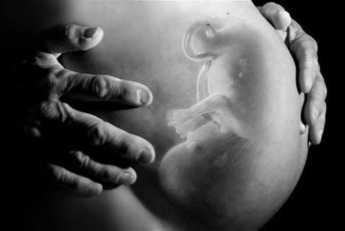
Women do not take X-rays for herniated discs while they are pregnant. pregnancy because it can affect the development of the fetus. Remove objects and equipment such as hairpins, jewelry, ... because they may affect the results of X-ray. Patients should go to the hospital with rheumatology to perform X-ray methods, to help detect the disease in time and have a quick treatment for herniated disc.

Please dial HOTLINE for more information or register for an appointment HERE. Download MyVinmec app to make appointments faster and to manage your bookings easily.
SEE MORE
Treatments for herniated discs Herniated discs: When to practice therapy, when to have surgery? Treating herniated discs with spinal manipulation at Vinmec Times City: Effective solution, without drugs


