This is an automatically translated article.
The article is expertly consulted by Master, Doctor Luu Thi Bich Ngoc - Doctor of Radiology - Department of Diagnostic Imaging and Nuclear Medicine - Vinmec Times City International General Hospital.Diffuse cranial magnetic resonance imaging is a valuable technique that provides information on the physiological status of the brain and is particularly sensitive to ischemic infarction. It is also recommended when patients have acute neurological deficits, traumatic brain injury, diagnose acute stroke and differentiate stroke from other diseases.
1. What is cranial diffusion magnetic resonance imaging?
Diffusion imaging (DWI) provides unique information on brain parenchymal viability. The sequences of T2W, FLAIR... are high signal pulses representing increased signal in the blood supply region of the middle cerebral artery. This pulse sequence is often imaged specifically in conjunction with diffusion pulses (DW) to evaluate the diffusion of water molecules in the lesion (often significantly variable by pathology), to predict the survival of the patient. brain parenchyma.Because diffusion imaging uses echo-planar imaging, it has little effect on patient movement and the survey time is fast, from a few seconds to a few minutes. DWI plays an important role in detecting cerebral infarction and distinguishing acute ischemic stroke from other intracranial pathologies.
2. Indication case
● Cerebrovascular accident (especially early cerebral infarction).● White matter diseases such as: scattered fibrosis, blood vessels, radiation...
● Inflammatory lesions of the brain, brain abscesses.
● Diagnosis of intracranial diseases (intra-axial, extra-axial brain tumors, neuromas, cystic lesions...).
● Traumatic brain injury, especially axonal damage....
● As prescribed by the treating doctor.

Người bệnh có bệnh lý u não cần được chụp cộng hưởng từ khuếch tán sọ não
3. Contraindications
● Patients wear electronic devices such as: cochlear implant, pacemaker, anti-vibration machine, Neurostimulator, automatic drug injection device under the skin....● Wear metal surgical clips Intracranial, orbital, vascular less than 6 months (If worn for more than 6 months, consider adding depending on the patient's condition).
● People with serious illnesses need resuscitation equipment on their side.
4. Brain diffusion magnetic resonance imaging procedure
4.1 Preparation before the procedure To perform cranial diffusion magnetic resonance imaging, it is necessary to prepare:Implementation team:
● Radiology specialist.
Electro-optical technician.
Vehicles used:
Resonant angiography machine from 1.5 Tesla or more.
Film printer.
Movies.
● Image storage system.

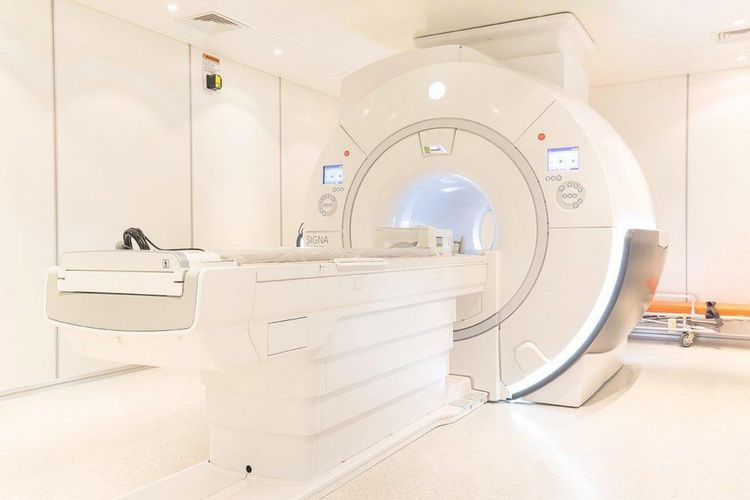
Máy chụp cộng hưởng từ khuếch tán sọ não yêu cầu từ 1.5 Tesla trở lên.
● No need to fast.
● Prepare a copy of the request from a clinician, with a clear diagnosis or complete medical record (if necessary).
● The procedure is explained in advance to coordinate well with the doctor.
● Check for contraindications.
● Change the clothes of the MRI room according to the instructions, remove the contraindicated items.
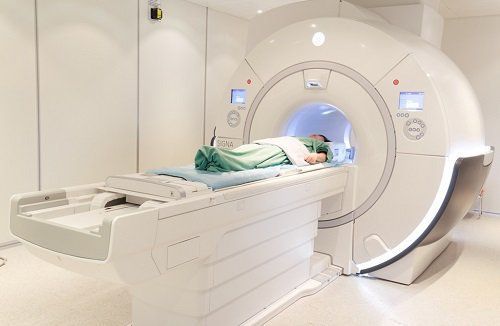
4.2 Procedure The whole procedure of cranial diffusion magnetic resonance imaging takes about 30 minutes in turn, following these steps:
Patient position:
● The patient is instructed to lie supine on the imaging table.
Select and position the receiver coil.
● Move the table into the machine bay and locate the shooting area.
Shooting techniques
● Positioning shooting.
● Take conventional T1, T2, FLAIR cranial pulse sequences first.
Capture the diffuse pulse sequences and the “b” value from b0 to b1000 depending on the specific clinical case.
Graph the apparent diffusion coefficient (ADC).
Evaluation of results
Based on the obtained image results, the doctor can assess the brain parenchymal damage (if any), the degree of brain parenchymal diffusion... from which to give expert judgment and advice training for the patient and the patient's family if required.
Note:
● During the scan: continuously monitor pulse, blood pressure and focal neurological signs.
● After the scan: let the patient wait for about 15 minutes for further monitoring or transfer to the emergency room.

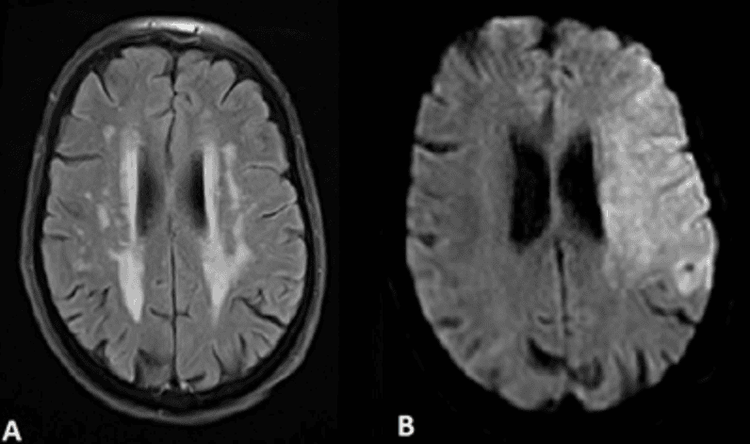
Chụp cộng hưởng từ khuếch tán sọ não trục cho thấy cường độ tín hiệu cao (B)
5. The role of cranial diffusion magnetic resonance imaging
Diffusion of the brain (DWI) will provide a different contrast than conventional MRI techniques. Diffuse cranial magnetic resonance imaging (MRI) is particularly sensitive for detecting acute stroke and distinguishing it from other conditions presenting with sudden neurological deficits.Diffuse imaging also provides supportive information for other brain pathologies such as brain tumors, intracranial infections, and traumatic brain injury. ... Because stroke is common and is the differential diagnosis of most acute neurological situations, diffusion imaging should be adopted as the essential pulse sequence and is recommended for most MRI investigations. from the brain.
Vinmec International General Hospital is a high-quality medical facility in Vietnam with a team of highly qualified medical professionals, well-trained, domestic and foreign, and experienced.
A system of modern and advanced medical equipment, possessing many of the best machines in the world, helping to detect many difficult and dangerous diseases in a short time, supporting the diagnosis and treatment of doctors the most effective. The hospital space is designed according to 5-star hotel standards, giving patients comfort, friendliness and peace of mind.
Master, Doctor Luu Thi Bich Ngoc has 06 years of experience working in the field of diagnostic imaging, received formal training and graduated with a Master's degree from Hanoi Medical University. Dr. Ngoc has strengths in diagnosing breast, thyroid, liver cancers early. experience in doing ultrasound of tissue elastography for liver, breast, thyroid diseases... From 2019 until now, Dr. Ngoc has worked at Vinmec Times City International Hospital with a position at the department. Image analysation.
To register for an examination at Vinmec International General Hospital, you can contact the nationwide Vinmec Health System Hotline, or register online HERE.


