This is an automatically translated article.
The article was written by Mr. BS. Bui Thi Hong Khang - Pathologist - Laboratory Department - Vinmec Central Park International General HospitalThe common cell types in benign effusions are mainly mesenchymal cells, macrophages (histocytes) and lymphocytes.
A. Cell composition
1. Mesothelial cells
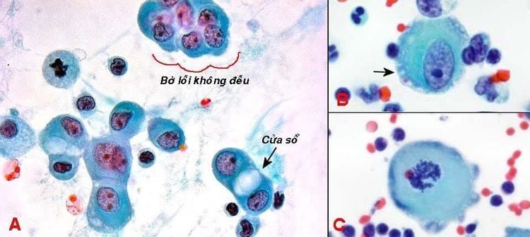
1.1 Normal mesothelial cells Normal mesothelial cells on histology section are flattened, when detached into the body cavity, they will be round, variable in size and located separately or in small plaques 2. dimension (less than 15 cells) with irregular convex margins. The cytoplasm is basophilic with the central region near the nucleus becoming darker than the periphery, and the cell border is quite clear or indistinct with the addition of an outer lace border (formed by microvilli).The nucleus is round or oval, the diameter of the nucleus is half the diameter of the cell, in the center or slightly eccentric, the chromatin is fine-grained, with 1 small nucleus, the nuclear membrane is well-defined. Between the two adjacent mesenchymal cells, a space called the window can be seen. Cell division is rare, but if seen, it does not mean cancer cells. (Figure 1)
1.2 Reactive mesothelial cells and degenerated mesothelial cells In effusions due to non-cancerous conditions (pulmonary infarction, heart failure, cirrhosis, renal failure) chronic, autoimmune disease ...), mesothelial cells are stimulated to proliferate into enlarged, square-shaped, nucleated cells; can be layered, even papillae. When degenerated, cells have many vacuoles in the cytoplasm, which are clearly seen on histological sections. (Figure 2)

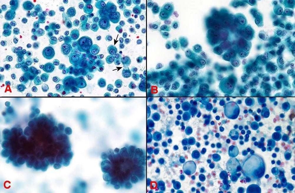
Figure 2: Proliferating pleural mesothelial cells with enlarged, square nuclei (A); can be arranged in many layers (B), papillae (C), when degraded, the cytoplasm has many vacuoles (D).
The smears from these benign effusions will have a high cell density (cell rich smears), reactive mesenchymal cells are mostly isolated but can also form small 2-dimensional clumps (less than 15 cells).
Cells and nuclei are irregular in size, have 1 or 2-3 nuclei, nucleus is still centrally or slightly eccentric, no hyperchromic, chromatin is still quite smooth, nucleus is larger and nuclear membrane remains uniformly clear, the nucleus/cytoplasmic ratio may be increased, but lace pellets and windows are still found (Figure 3A). The mitotic rate was increased but there was no abnormal mitotic pattern.
Reactive mesothelial cells may form spherical, 3-dimensional clumps resembling mulberry fruit (morula), glandular sacs or papillae; Although the clusters are usually small in size, the papillae are not branched, but can sometimes be mistaken for mesothelioma. (Figure 3B,C)
| Calretinin | EMA | Desmin | GLUT-1 | p53 | |
| Tế bào trung mạc phản ứng | + | - | + | - | - |
| Tế bào trung mạc ác tính | + | + | - | + | + |
Table 1: Distinguishing benign and malignant mesothelial cells by immunocytochemistry
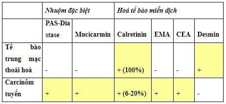
When mesothelial cells are degenerated, the cytoplasm has many glycogen-containing vacuoles, causing the nucleus to be displaced to one side. may be mistaken for mucin-secreting cancer cells. Distinguish by special staining or immunocytochemistry (Table 2)
1.3 Histiocytes / Macrophages Are the type of cells that are always found in isolated effusions. The nucleus is oval or kidney-shaped, lying on one side, the chromatin is sparse, the nucleus is usually small; cytoplasm is abundant and pale, with many vacuoles that can contain phagocytic objects (dead lymphocytes, nuclear debris...). (Figure 4A)
1.4 Lymphocytes Found readily in effusions, the majority are T lymphocytes. Cells are small in size, round nuclei are dark purple, nuclei are not clear, and there is very little cytoplasm. (Figure 4B)
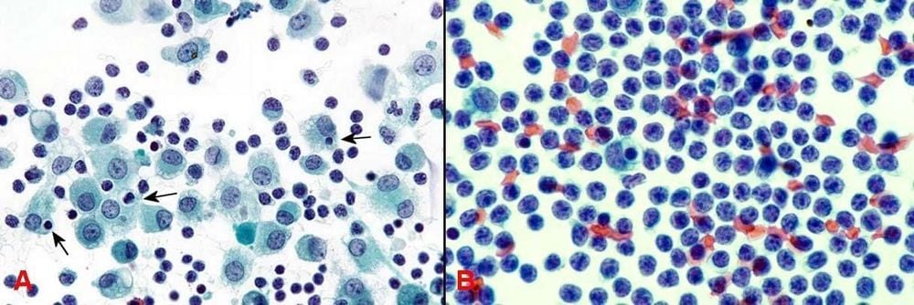
In addition to the 3 main cell types, the mesenchymal cells, macrophages (histocytes) and lymphocytes; In the effusion, other cell types such as neutrophils, eosinophils, and red blood cells can also be seen.
B. Some types of effusion in benign diseases
1. Purulent effusions
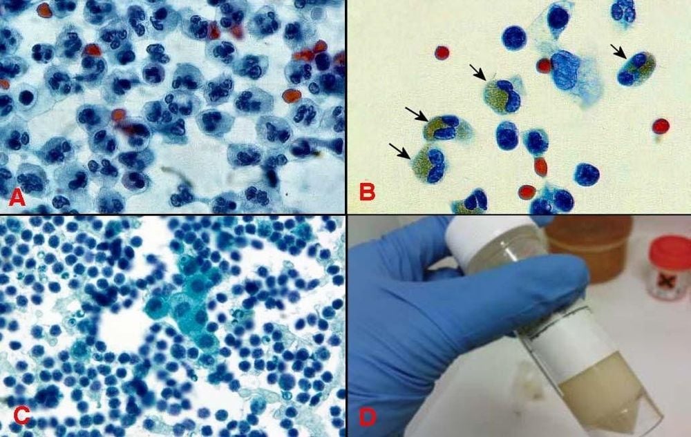
Bacteria can cause acute inflammation of the pleura, peritoneum and pericardium by various mechanisms, leading to pus overflow in the body cavity. The pus is pale yellow or greenish in color and may have a foul odor. The smear showed mainly neutrophils, dead cell debris, some macrophages and lymphocytes; mesenchymal cells were rarely seen.
2. Eosinophilic Effusions
It is called an eosinophilic effusion when the number of these cells makes up more than 10% of the nucleated cells. Eosinophilic effusion can occur when the body cavity is repeatedly aspirated, allowing blood and gas to enter it; or due to parasitic infection, drug reaction, pulmonary infarction. The smear is rich in eosinophils, the nucleus is 2 lobes, and the cytoplasm is filled with orange granules.
3. Lymphocytic effusions
It is called a lymphocytic effusion when the number of these cells accounts for more than 50% of the nucleated cells. May be due to tuberculosis, collagen disease or after a coronary bypass surgery. Clear, pale yellow, sometimes slightly cloudy, and bloody discharge. The cytology consists of small lymphocytes, mesenchymal cells and histiocytosis.
4. Chyous effusions
Rarely, occurs due to trauma or surgery to rupture the thoracic duct causing the chyme to overflow into the body cavity. Chylous effusion is milky white because it contains a lot of lipids and a few lymphocytes.
It should be noted that all of the above types of effusions can also occur in the setting of cancer; Therefore, caution should always be exercised to look for malignancies when reading cytological specimens.
Please dial HOTLINE for more information or register for an appointment HERE. Download MyVinmec app to make appointments faster and to manage your bookings easily.
References source: Textbook of cytology Pham Ngoc Thach Medical University


