This is an automatically translated article.
The article was written by Mr. BS. Bui Thi Hong Khang - Pathologist - Laboratory Department - Vinmec Central Park International General HospitalWhen a patient has pleural, pericardial, or peritoneal effusion, doctors usually test for pleural, pericardial, or peritoneal fluid and cell block.
1. Classification of effusion
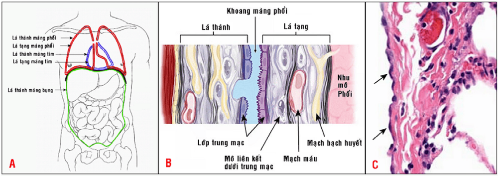
In humans there are 3 main body cavities: the pleural cavity, the peritoneal cavity and the pericardial cavity. The body cavity is lined by a layer of flattened mesothelial cells, resting on a layer of connective tissue containing collagen fibers, elastic fibers, many blood vessels and lymphatic vessels and nerve fibers; forms the visceral pleura (which surrounds the lungs, gastrointestinal tract, and heart) and parietal (applies to the chest wall, abdominal wall, and mediastinum). (Figure 1)
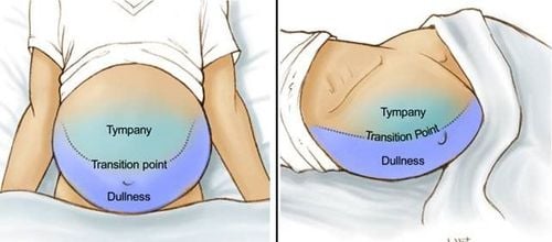
It is the balance between these factors that helps to maintain the amount of fluid in the body cavity only a few milliliters, while the amount of fluid produced per day can be up to 5-10 liters; so, when this imbalance is lost, fluid will pool in the body cavity, causing effusion (fluid in the peritoneal cavity also known as ascites).
Two types of fluid can be distinguished - transudative and exudative - based on their distinct characteristics as shown in (table 1).
| Dịch thấm | Dịch xuất | |
| Tỷ trọng | <1,012 | >1,020 |
| Protein | < 3 g% | ≥ 3 g% |
| Đại thể | Trong, màu vàng lợt | Đục, màu sắc thay đổi |
| Tế bào | Vài tế bào trung mạc và bạch cầu | Giàu tế bào (>300 tế bào /mm3 ) |
| Nguyên nhân |
Suy tim, Suy thận ; Xơ gan, Hội chứng thận hư, Suy dinh dưỡng |
Bệnh lý viêm nhiễm ; U nguyên phát hoặc di căn |
| Cơ chế bệnh sinh | Tăng áp lực thủy tĩnh ; Giảm áp lực thẩm thấu keo | Tăng tính thấm thành mạch ; Giảm dẫn lưu vào mạch bạch huyết |
2. Body fluid cytology technique
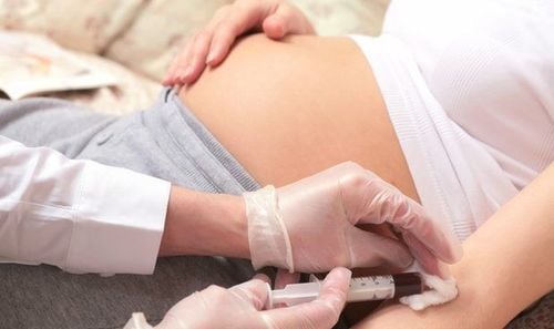
Body cavity fluid is removed by needle aspiration or drainage for both therapeutic purposes (decompression) and diagnostic sampling (Figure 2).For cell-rich fluid samples, can be smeared directly on the slide; If there are not enough cells, centrifuge, take the sediment and spread it on a slide, the remaining residue is molded into a cell block for thin cutting (these thin slices are very convenient for performing other methods). special staining and immunocytochemistry).
For fluid samples containing a lot of blood, density gradient centrifugation can be used to separate cells of interest.
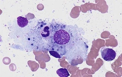
Cytological slides were fixed wet or dry and stained according to the Papanicolaou or May-Grunwald-Giemsa method.

Please dial HOTLINE for more information or register for an appointment HERE. Download MyVinmec app to make appointments faster and to manage your bookings easily.
References: Textbook of Cytology of Pham Ngoc Thach Medical University


