This is an automatically translated article.
The article is professionally consulted by Master, Doctor Le Xuan Thiep - Department of Diagnostic Imaging - Vinmec Ha Long International Hospital
Biliary surgery in hepatobiliary ultrasound is a safe, simple technique but plays an important role in locating extrahepatic biliary tract lesions, thereby helping doctors detect abnormalities and lesions. liver, bile.
1. What is hepatobiliary ultrasound?
Ultrasound is an imaging technique that uses high-frequency sound waves (ultrasound waves) to shape and reconstruct images and structures inside the body. This is one of the paraclinical methods to diagnose, monitor and treat common diseases, applied in most medical facilities.
Hepatobiliary sonography is an ultrasound of the liver and biliary tract that can identify the lobes, segments, and subsegments of the liver, based on the relationship of the inferior vena cava, aorta, hepatic vein, and vena cava. door.
2. Structure of the bile ducts inside and outside the liver
The gallbladder is a small, green, pear-shaped sac located on the underside of the right lobe of the liver, about 6 to 8 cm long and 3 cm wide when fully distended, containing about 30-50cc of bile. This is a part of the extrahepatic bile duct, which consists of 3 parts: base, body and neck.The bile duct from the gallbladder (cystic duct) to the common bile duct is 3-4cm long, 4-5mm wide at the beginning, 2.5mm narrow at the end. In the upper part of the cystic duct, there are valves called Heister valves that keep the cystic duct from folding and allow bile to flow easily.
The gallbladder and bile duct system are nourished by the branch of the hepatic artery, which is the direct connection between the liver and the gastrointestinal tract. The gallbladder is cone-shaped, 7-10cm long, containing 30-50cc of bile (repeated). From each lobe of the liver, bile from the small ducts empties into a common duct, and then into the common hepatic duct leading to the gallbladder. Bile from the pouch follows the common bile duct into the duodenum in the ampulla of Vater. There are 60 - 70% of adults with pancreatic duct emptied into the common bile duct before entering the duodenum, so acute cholecystitis (may be related to or lead to acute pancreatitis, especially due to obstruction: stones, . ...) is associated with pancreatic necrosis.
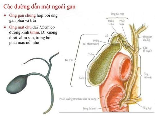
Hình ảnh túi mật và hệ thống ống mật
3. What is the reserve function of the gallbladder?
A functioning liver produces bile continuously. Bile is a slightly thick substance with a greenish-yellow color and a bitter taste. Bile plays an important role in the digestion of food. Bile salts include Sodium Glycocholate and Sodium Taurocholate that are responsible for breaking down fats, promoting the activity of Lipase enzymes to break down Lipids. They also help digested fats to pass through the intestinal wall.
4. Anatomy of the biliary tract
Biliary tract includes intrahepatic and extrahepatic bile ducts.
The intrahepatic bile ducts originate from the intralobular tubules and then empty into the perilobular ducts. These pipes are joined together and coalesce at the opening to form larger pipes. The location of the intrahepatic bile duct resembles that of the portal vein. Each segmental vein has one or two bile ducts that enter the hilum of the liver to form a right hepatic duct and a left hepatic duct.
The extrahepatic biliary tract (main biliary tract) has 4 segments: the hilum, the omentum, the posterior pancreaticoduodenal and the intramural. The first two segments contribute to the hepatic peduncle, which consists of the major components that travel to and from the liver via the hilum: the portal vein, the hepatic artery, and the major bile duct (the common hepatic duct). In particular, the portal vein is located in the posterior plane of the hepatic peduncle, the anterior plane consists of the descending main bile duct on the right and ascending hepatic artery on the left.
5. Hepatobiliary ultrasound
5.1 Hepatobiliary ultrasound method: transabdominal ultrasound Before the ultrasound, the patient will be asked to fast for 6-8 hours, because if eating, the gallbladder will shrink and make it difficult to examine, leading to to miss minor injuries. However, in emergency cases, the doctor can perform an ultrasound but still need to do a clinical examination.
5.2 Procedure for performing hepatobiliary ultrasound The patient lies in the supine position, inhales deeply and holds the breath in order to lower the liver and avoid gas in the colon. The doctor applies a gel to the abdomen with the effect of helping the detector make sure contact with the body, avoiding the air between the detector and the human skin.
Perform ultrasound of the entire abdomen including the area around the liver - bile.
>>> The role of ultrasound, computed tomography in diagnosis and treatment of hepatobiliary disease
After the end, the doctor will wipe the gel that was initially applied and end the ultrasound process. Patient waits for results according to instructions.
Intrahepatic bile ducts are difficult to see if they are not dilated. The main biliary tract analyzed on diagonal or parallel layers below the ribs, it is a tubular structure located in front of the portal vein, usually less than 7mm in diameter, may be larger after surgery or in the elderly.

Siêu âm gan mật được thực hiện tại vùng bụng của bệnh nhân
In the case of obstructive dilatation, dilated intrahepatic biliary tract manifests as hypoechoic tubules in the liver parenchyma resembling a “crab foot” or a “spider leg”, converging to the hilum, anterior to the branches of the veins. door. When the extrahepatic bile duct is obstructed, the left hepatic duct usually dilates earlier than the right hepatic duct.
Dilated extrahepatic biliary tract, seen on subcostal sections (transverse or retrograde), is a "double-lumen gun" shape in the hepatic pedicle (posterior portal vein and anterior common bile duct), which should be distinguished from pseudoform formed by the cystic duct or hepatic artery with the portal vein.
Please dial HOTLINE for more information or register for an appointment HERE. Download MyVinmec app to make appointments faster and to manage your bookings easily.