This is an automatically translated article.
The article is expertly consulted by Master, Doctor Hoang Van Lan Duc - Doctor of Radiology - Department of Diagnostic Imaging and Nuclear Medicine - Vinmec Times City International General Hospital. The doctor has more than 10 years of experience in the field of imaging, especially in diagnosing with emergency internal, surgical, abdominal, thoracic, musculoskeletal, neurological and thyroid, breast, ...Chest X-ray is a technique used to diagnose the patient's lungs to evaluate the condition of the lungs, its components and nearby structures.
1. Basic lesions in lung disease detected on X-ray
X-ray is the most used imaging technique in medicine, helping doctors to evaluate and detect abnormal symptoms of chest X-ray films, thereby helping clinicians make informed decisions. preliminary diagnosis. The article will provide information about the basic lesions of the lungs detected on radiographs.Based on the degree of X-ray blocking of the lesion, lung lesions are divided into three types: watermark, bright image and combined watermark - light.

2. Blurred lung lesions
Depending on the size, opacities are divided into 3 types: opacity of a lung field, opacities, opacities, and opacities.2.1 Blur an entire waste field:
Common in the following diseases:Complete effusion on one side of the pleural cavity.
The cause may be tuberculosis, lung cancer or trauma...
Solid image of the entire lung, pushing the surrounding organs. The damaged intercostal space is enlarged, the mediastinum is pushed towards the healthy lung,...
Thickening of the entire pleural cavity.
Often due to sequelae of poorly treated pleural effusion, hemothorax due to trauma, ..
Solid image of the entire lung, causing a very strong pulling effect. Pulling the mediastinum toward the injured side, causing the intercostal space on the injured side to narrow. Pleural calcifications can be seen on film.
Total collapse of one lung.
Due to obstruction of the main bronchus. The most common cause is a patient with central bronchial cancer or a foreign body in the main bronchus.
A watermark appears on the entire lung causing a tug-of-war effect. However, the degree of tensile strength is usually much lighter than that of the adhesive.
2.2 Clouds:
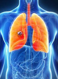
Used to refer to watermarks that are about 3-10cm in size. May occupy one lobe or one lobe of the lung. The opacities are common in the following diseases:Pleural effusion: The opacities are homogeneous in the lung base and the costophrenic angle, the upper boundary is not clear, forming the Damoiseau curve. Pleural effusion causes compression effect depending on the degree: push mediastinum to the opposite side, dilate the intercostal space,... Lobar pneumonia (common pneumococcal pneumonia): Most commonly in the middle lobe right lung. At the typical stage of the lesion, there is a homogeneous opacity in the lower half of the right lung, the right flank angle is bright, the upper border is clear, and the small interlobular fissure is present. On tilt film, the opacities have a triangular shape occupying the entire right middle lobe of the right lung, the edges are straight or convex, the apex turns into the hilum. The two edges are two interlobar grooves. Atelectasis: The opacities are relatively homogeneous in the form of a triangle with concave sides. If one lobe or segment collapses, the opacity is an interlobar fissure. Common in bronchial Adhesions Pleural thickening : As a consequence of pleural effusion. The cloud is homogeneous or relatively homogeneous, the boundaries are often clear. Causes contraction of related organs (pharyngeal arch, mediastinum, intercostal space, ..).

+ Peripheral bronchial cancer is located in the periphery of the lung. Because the malignancy of peripheral lung tumors is usually low, the tumor appears to be a benign mass. Round or oval in shape, smooth edges, often well-defined boundaries.
Tuberculosis : The cloud is not homogeneous, the boundary is not clear. Lesions at different stages of development: infiltration, fibrosis, calcification, ..
2.3 Faded notes:
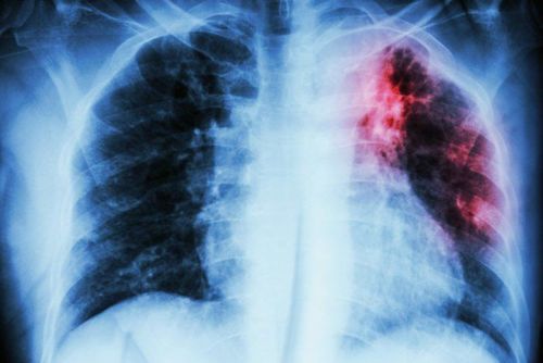
These are watermarks that are 1-3cm in size. Spotted lesions are common in the following diseases:Cancer metastasis to the lung: Round, or oval, opacities, unequal in size, have the appearance of dropping balloons scattered throughout the two lung fields.
Tuberculosis lesions:
Tuberculosis infiltrates: Non-homogeneous opacities often in the apex, subclavian region, unclear boundaries, at different stages. Tuberculosis: A solitary hazy spot in the periphery of the lung, usually round, with smooth margins, well-defined boundaries, possibly with calcifications.
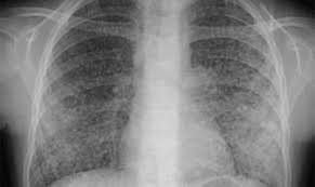
2.4 Small millet faint:
These are opacities ranging in size from 1 to 10 mm, commonly seen in the following diseases:TB: Tiny opacities, about 1 mm in size, evenly distributed throughout the two lung fields. Migraine cancer metastasis: Small and irregular opacities like in millet tuberculosis, concentrated mainly in the umbilicus and lung base.
3. Light image
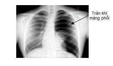
Pneumothorax:Bright image is usually in the periphery and upper part of the lung, no pulmonary striations, limited with the lung parenchyma as the border of the visceral pleura. The lung parenchyma is pushed toward the hilum (passive atelectasis). Causes pushing effect depending on the degree (push mediastinum to the healthy lung, dilation of the intercostal space,...).
Cocoon:
Light shape is usually round or oval, thin wall, smooth border, well demarcated. Air cysts are usually solitary on one lung, with different sizes. The nature of the air cocoon is loss of pulmonary ridges. However, because radiographs are the result of anterior-posterior superposition, only large air cysts occupying the entire anteroposterior thickness of the chest wall are absent. Smaller air cysts are superimposed by the healthy lung parenchyma before and after it, so the striae are still visible.
Tuberculosis:
A bright image located on an area of the lung with surrounding TB lesions. The cave wall is often thick, the border is not clear, there are many infiltrative lesions around. Old caves often have thin walls, smooth edges, and clear boundaries. Around there are many fibrous bands, calcified nodules. Because of the old TB lesions, there is often a tug-of-war effect due to thickening of the pleura.
4. Watermark - light combination
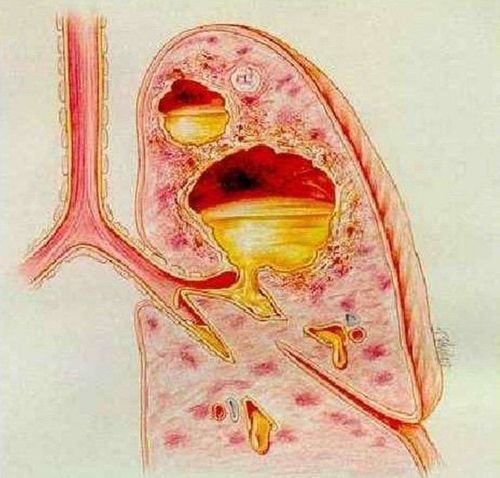
Lung abscess:Chest radiograph shows round or oval appearance, well demarcated, often solitary in lung abscess with direct spread or multiple small foci in the case of pulmonary abscess caused by staphylococci bacteria. In the purulent stage, the cavern is formed with gas and fluid levels. These are masses located in the lung parenchyma, with thick crust and clearly demarcated with healthy lung tissue. Light image is on top, lung ridges are lost. Solid watermark at the bottom. Bright and fuzzy image boundaries are horizontal levels. This pattern should be distinguished from an infected air cocoon. The cocoon wall is usually thin, and the fluid level is low. The wall of the abscess is thick, and the fluid level is high.
Pneumothorax, pleural effusion:
Lesions are located in the periphery of the lung, the location of the pleural cavity. The light image has lost the pulmonary ridge above, pushing the parenchyma into the hilar area. The bottom solid watermark creates a horizontal level.
It can be seen that, with abnormalities appearing when taking a chest X-ray, the doctor can detect and diagnose the underlying lesion in lung disease based on his signs and experiences.
Results of the patient's examination will be returned to the home. After receiving the results of the general health examination, if you detect diseases that require intensive examination and treatment, you can use services from other specialties at the Hospital with quality treatment and services. outstanding customer service.
Please dial HOTLINE for more information or register for an appointment HERE. Download MyVinmec app to make appointments faster and to manage your bookings easily.


