This is an automatically translated article.
The article was professionally consulted by BSCK II Nguyen Quoc Viet - Interventional Cardiologist - Department of Medical Examination & Internal Medicine - Vinmec Danang International General Hospital.In cardiovascular disease, endovascular intervention is increasingly being used worldwide. Success in this field is attributed to the extremely important contribution of digital background angiography (DSA). Strictly speaking, DSA is a golden procedure in the imaging of cardiovascular diseases in the world.
1. Cardiovascular background removal digital angiography
Digital Subtraction Angiography (DSA) is a new X-ray angiography system. Digital Subtraction Angiography (DSA) is an imaging method that combines X-ray imaging. and digital processing using algorithms to remove the background on 2 images obtained before and after the contrast agent is injected into the patient's body, obtaining a full image of the heart and carotid, thoracic, abdomen and arteries thigh was obtained, in order to study the blood vessels in the body and better see the lesions and vascular pathology before vascular intervention is indicated. This is the product of a combination of computerized image processing techniques and conventional angiography by Seldinger technique.The components of a DSA system typically include an X-ray generator, an image acquisition unit, a digital image processor, and a display unit.... The central part of a DSA system is the digital image processing system. Before performing digital background erasure angiography, a fluorescent dye is injected into the patient. This contrast agent has the effect of lightening blood vessels, this dye is completely harmless and will be excreted from the body through the patient's urine. The X-ray is then emitted, and passes through the patient's body and is picked up by the Image Intensifier. A lens is placed between the bulb and the video camera for the purpose of limiting the intensity of light that reaches the camera. Then, the blood vessel to be imaged is injected with contrast material through a catheter that is inserted into the femoral artery through the skin. When the contrast material enters the body, the machine will capture the animation in 1 preset time unit. When there is no contrast agent as the background image, the image processing unit will take the acquired image (mask image) and proceed to exclude the background image with the image obtained when the contrast is present, which are static anatomical structures. similarity between the two images.
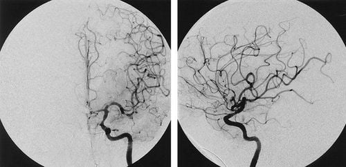
Hình ảnh chụp động mạch bằng kỹ thuật chụp mạch máu số hóa xóa nền
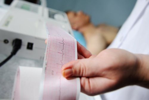
Before background digitized angiography, the patient must be carefully prepared including tests such as: recording electrocardiogram, blood test, chest X-ray, and the patient must fast for breakfast. Digital angiography with background erasure is an invasive imaging technique and carries a risk of complications when taking pictures. Therefore, it must be strictly prescribed and must be agreed to by the patient. After the scan, the patient will be bandaged in the groin area and lying motionless for 24 hours, and absolutely avoiding leg movement.
2. Advantages of DSA . technique
The advantage of this technique is that it helps doctors to detect early and accurately abnormal blood flow conditions, which helps in the accurate diagnosis and treatment of serious diseases related to blood circulation. in the body, planning the anatomy and helping to pinpoint the exact locations of damage inside the body. Thanks to this DSA machine, quite a few cases of cardiovascular disease, especially myocardial infarction, were clearly diagnosed and were intervened quickly and within the golden period, so their lives were saved.
Hệ thống chụp mạch số hóa xóa nền DSA tại Bệnh viện Đa khoa Quốc tế Vinmec
Digital angiography erasing DSA background in cardiovascular disease has been widely used. At Vinmec International General Hospital, there is a team of experienced specialists, along with modern equipment, using digital angiography to erase the background of DSA in the heart, helping doctors in the diagnosis. and the treatment is quick and accurate.
Doctor Nguyen Quoc Viet has more than 20 years of experience in the examination and treatment of cardiovascular diseases and Cardiovascular Interventions (Including angiography, dilation, stenting of coronary arteries, renal arteries...), placing temporary, permanent pacemaker... Before working at the Department of Medical Examination & Internal Medicine, Vinmec International General Hospital Da Nang, the doctor used to work for a long time at Da Nang Hospital.
Please dial HOTLINE for more information or register for an appointment HERE. Download MyVinmec app to make appointments faster and to manage your bookings easily.