This is an automatically translated article.
The article was professionally consulted with Specialist Doctor II Nguyen Quoc Viet - Department of Medical Examination & Internal Medicine - Vinmec Da Nang International General Hospital.Acute aortic arch syndrome is a potentially life-threatening condition with initial clinical presentation within 14 days. Because blood pressure in the aorta is high, it is necessary to quickly diagnose and treat in time to preserve the patient's life.
1. What is acute aortic arch syndrome?
Acute aortic arch syndromes are disorders of the thoracic and abdominal aorta with symptoms that require urgent evaluation and review, sometimes requiring surgical intervention.This group of pathologies includes acute aortic dissection, intramural hematoma, and infiltrative atherosclerotic plaque ulceration. Approaching diagnosis and treatment of these conditions often requires multi-specialty coordination, including family physicians and internal medicine, emergency medicine, critical care, imaging, cardiology, and surgery heart surgery.
2. Acute aortic dissection
2.1. What is acute aortic dissection? Acute aortic dissection is the most common as well as the most fatal condition of acute aortic syndrome, and therefore requires urgent diagnosis and treatment. If left untreated, this condition increases mortality by 1% to 2% per hour after the onset of symptoms and 90% at three months.Aortic dissection results from a tear in the aortic wall and blood enters the medial layer of the aorta. This mechanism divides the aorta into true lumen and false lumen, which is separated by a septum from the aortic medial layer. As the outer wall of the aorta has become thinner and less supportive, a sudden high intravascular pressure can cause rupture of the blood vessel, spillage of blood into the pericardial space, and fatal cardiac tamponade. . Therefore, the size of the dissected hematoma determines the outcome. Sometimes the location of the hematoma originates from a tear in the proximal ascending aorta, extending far away, which can block important aortic branches, such as the arteries that supply blood to the brain and kidneys. , abdominal viscera and extremities. If the hematoma spreads posteriorly, the coronary artery can reduce blood supply, leading to myocardial infarction, or severe acute aortic regurgitation, causing heart failure.
2.2. Risk factors for aortic dissection Hypertension is the most common risk factor for acute aortic dissection. Next, severe mediastinal degeneration may be the cause in about 20% of patients. Other risk factors include the presence of inherited connective tissue abnormalities, mitral valve prolapse, ascending aortic dilatation, aortic root, and aortic spasm. In addition, cocaine use, catheter interventions into the aorta and coronary arteries, and manipulations during open-heart surgery can also lead to aortic dissection.
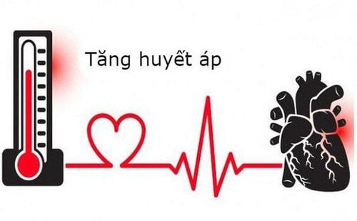
Tăng huyết áp là yếu tố nguy cơ phổ biến của hội chứng cung động mạch chủ
Stanford class A includes lesions occurring at any anatomical site involving the ascending aorta, with or without involvement of the aortic arch and distal aorta.
Stanford class B includes lesions occurring at any anatomical site in relation to the descending thoracic aorta, which can sometimes extend proximally to the aortic arch.
In addition, acute aortic dissection can also be classified according to DeBakey, type I is similar to Stanford A and type III is similar to Stanford B. DeBakey II type is the type of lesion that originates and is limited to ascending aorta and is the least common type.
2.4. Symptoms of aortic dissection The most common symptom of acute aortic dissection is sudden and severe chest pain. However, other variants of chest and back pain have been described and acute aortic dissection can also occur in the absence of pain.
In Stanford type A, the pain is usually located in the anterior chest area and may radiate to the neck, back, or abdomen. In Stanford type B, the pain usually occurs behind the chest and also in the abdomen.
Other possible symptoms are syncope due to hypotension related to cardiac tamponade or rupture of the aorta; coma due to severe blockage of a cerebral artery; abdominal pain may also occur due to decreased perfusion of the mesenteric and renal arteries; Renal failure during acute aortic dissection has a mortality rate of 50-70% and mesenteric ischemia with a mortality rate as high as 87%. If palpation is difficult in the legs, the possibility of thoracic aortic dissection should be considered with occlusion of the iliac and abdominal aorta.
2.5. Diagnosis Patients presenting to the emergency department with significant chest pain will undergo an electrocardiogram and serum troponin measurement to rule out myocardial infarction. However, it is important to note that the ECG can be abnormal in up to 70% of patients with ascending aortic dissection. Therefore, if myocardial infarction is suspected, other diagnostic tools should also be performed immediately to exclude aortic dissection, in order to avoid the use of drugs that affect coagulation.
Chest radiographs may show enlarged upper mediastinum, but are usually insignificant. Meanwhile, contrast-enhanced computed tomography is currently the most widely used diagnostic tool to detect aortic dissection.
Transesophageal echocardiography has high sensitivity and specificity for detecting dissection of the ascending aorta, which is superior to transthoracic echocardiography. This tool can be performed quickly but requires a sedation and a cardiologist to perform. In addition to determining the dissection layer, transesophageal echocardiography can identify pericardial tamponade, aortic regurgitation, and the location of the affected coronary artery origin. Furthermore, the location of the tear is also determined, which is very helpful for the surgeon. Meanwhile, transthoracic echocardiography has only limited value.

Đau ngực đột ngột là triệu chứng điển hình của hội chứng cung động mạch chủ
2.6. Treatment of aortic dissection The initial treatment of Stanford type A aortic dissection is rapid control of blood pressure and heart rate, thereby reducing the left ventricular contraction rate, preventing the spread of the mass. the hematoma is dissecting. Intravenous beta-blockers are used to slow the heart rate. The potential for reduction in afterload is achieved with intravenous administration of sodium nitroprusside, clevidipine or nicardipine. At the same time, the patient was also prepared for emergency surgery. The goal of surgery is to prevent death from pericardial tamponade or haemorrhage through removal of the dissection site by resection of the damaged aorta and replacement with a synthetic graft. blood flow into the true vessel.
For patients with Stanford type B aortic dissection, initial management is similar with beta-blockers, afterload reduction to control blood pressure and pain relief without surgery. Immediate surgical intervention is indicated only when there is evidence of dissection rupture or rapid expansion with increased aortic diameter, ischemia due to poor perfusion, hypertension, and pain due to aortic dissection. uncontrolled dissection.
3. Hematoma in the wall of the aorta
As an acute aortic disease, intra-aortic hematoma is usually caused by vascular rupture and hematoma develops in the aortic wall. Patients may present with symptoms and signs similar to aortic dissection. At this point, initial evaluation and management are beta-blockers and hypertension control.However, the natural course of hematoma in the aortic wall can vary from person to person. In which, if the diameter of the aorta is larger (> 5.5 cm) and the thickness of the hematoma increases rapidly (> 16 mm), the prognosis is poor. Thus, treatment of these subjects in the presence of severe chest pain or hemodynamic instability is the same as for patients with Stanford class A aortic dissection.
4. Penetrating atherosclerotic ulcer
Infiltrative atherosclerotic ulcer is a form of aortic arch syndrome, but it is less fatal than acute aortic dissection and intramural hematoma. However, patients may also present with severe chest pain that should still be carefully evaluated.In fact, infiltrative atherosclerotic ulcers are often discovered incidentally on computed tomography of the aorta or thoracic and abdominal magnetic resonance imaging performed for other indications. The site of injury usually occurs in dense calcified areas of atherosclerotic plaque in the thoracic aorta and diminishes as it descends into the abdominal aorta. A small percentage of ulcers will gradually enlarge, forming a pocket aneurysm or hematoma in the wall. At this point, surgical intervention may be indicated for endovascular repair and is preferred over open surgery.

Một dạng khác của hội chứng cung động mạch chủ là loét xơ vữa thâm nhập
5. Monitor for acute aortic arch syndrome
Patients who survive the acute phase of aortic disease, especially acute aortic dissection, should be closely monitored after initial therapy to detect aortic aneurysm formation in the early stages. residual artery, extension of dissection mass or intramembrane hematoma, as well as spread of penetrating ulcer.Computed tomography or magnetic resonance imaging and tight blood pressure control with beta-blockers or other antihypertensive agents are the mainstays of treatment and monitoring of these patients in the subacute and chronic stages. count. Any patient found to have a thoracic aortic aneurysm with a diameter of 4 cm or more should be treated appropriately and monitored periodically to determine when surgical intervention is needed. Untreated hypertension is a risk factor for aortic aneurysms and ruptures. Other risk factors, such as smoking and dyslipidemia, should also be controlled.
In cases where the patient is asymptomatic but the ascending aorta is 5.5 cm in diameter or larger or there is evidence of aortic dilatation more than 0.5 cm in one year should be considered for surgery. . The threshold for size criteria may be higher for aortic aneurysms involving the aortic arch and descending thoracic aorta while being lower in patients with genetic abnormalities.
In summary, acute aortic syndromes are disorders of the thoracic and abdominal aorta that are often symptomatic and require urgent evaluation and treatment. This group of pathologies includes acute aortic dissection, intramural hematoma, and infiltrative atherosclerotic plaque ulceration. The knowledge of history, presentation, timely diagnosis and proper surgical intervention are the “keys” leading to a successful outcome in aortic arch syndrome.
Please dial HOTLINE for more information or register for an appointment HERE. Download MyVinmec app to make appointments faster and to manage your bookings easily.