This is an automatically translated article.
Article written by Doctor Mai Vien Phuong - Department of Medical Examination & Internal Medicine - Vinmec Central Park International General Hospital
Celiac disease is also known as gluten sensitivity or gluten intolerance. This is an allergy to a form of protein called gluten, which does not allow the body to absorb gluten.
1. What is Celiac Disease (Gluten Intolerance)?
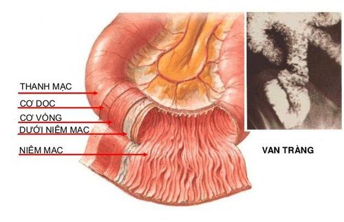
Gluten is found in many flours and grains, such as barley, wheat, and oats. This allergic disease affects mainly the small intestine, where food is stored after leaving the stomach.
2. What are the signs and symptoms of Celiac disease?
Common symptoms of Celiac disease include: diarrhea, accompanied by gray or liquid stools, often foul-smelling, oily and foamy looking; weight loss; growth and development delay (in young children); frequent bloating; abdominal swelling or pain; mouth sores; tired; weak people; Haggard; rash or muscle cramps.
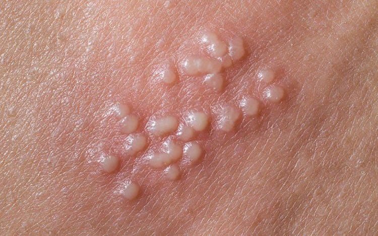
Adults with Celiac disease often have fewer symptoms than children, so your doctor will diagnose it with a blood test to detect abnormal anemia. Another rare manifestation of Celiac disease is herpes.
You may experience other symptoms and signs not mentioned. If you have any questions about the signs of illness, consult your doctor.
3. Causes of Celiac Disease
Guten plays an important role in the pathogenesis of Celiac . Gluten is the prolamin (plant protein) in grains including giadin in wheat, Sealin in rye, hordein in barley.
These are peptide chains rich in proline and glutamine, so they are difficult to break down by gastric acid, pancreatic enzymes or intestinal enzymes, even in healthy people. In normal humans, the epithelial layer of the intestinal mucosa does not allow passage of the wheat-derived gliadio peptide chains. Celiac is a chronic autoimmune disease caused by exposure to gluten in the lining of the digestive tract.
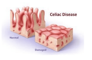
In this disease, the physiological protective barrier of intestinal epithelial cells is disrupted by giadin chains passing through the epithelial layer, activating a variety of immune mechanisms that damage intestinal epithelial cells. .
4.Role of innate and responsive immunity in Celiac disease
Damage in Celiac disease is fully reversible upon removal of gluten from the diet and reappears if the food contains gluten, Both the appearance of TG2 antibodies and an increase in the number of cytotoxic T cells in the epithelium are depends on and changes in the presence of gluten.
In patients with Celiac, there is an increased concentration of interleukins, mainly IL-15, and increased expression of group l histocompatibility complex molecules in the epithelium. All these factors suggest a strong response of the adaptive immune system as well as the involvement of the innate immune system in the complex pathogenesis of Celiac.
To summarize, gluten affects the intestinal mucosa through two mechanisms. The first mechanism is that the 19-mer degraded peptide fragments induce nonspecific innate immune responses that activate IL-15 production from enterocytes. This interleukin promotes the release of NF-KB and oxidants such as nitric oxide that activate programmed cell death as well as play a key role in "opening" the binding site between epithelial cells. tissue. The second mechanism is through an immune response that increases cell membrane permeability, allowing the 33-mer degraded peptide fragment to reach the subepithelial junction and be de-amide radicalized by the enzyme TG2. dendritic cell-activated IL-15 increases the expression and expression of gluten amide deprotonated peptides and activates a series of Thi-responsive pathways that release IFNY.
5.The role of endoscopy in the diagnosis of Celiac disease
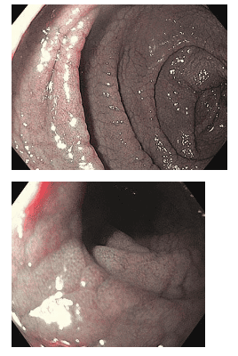
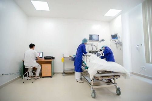
With the development of endoscopic techniques, more and more endoscopic images suggestive of Celiac disease have been recorded, thereby helping the endoscopic surgeon to conduct biopsies more easily.
Typical endoscopic images of the descending duodenum after inflation include: reduced number of duodenal mucosal folds (Kercking's fold), rough duodenal mucosa, visible network submucosa vascular network (mucosal atrophy), may show deep grooves, fissures, examination, papules, or lithographic images.
Please dial HOTLINE for more information or register for an appointment HERE. Download MyVinmec app to make appointments faster and to manage your bookings easily.


