This is an automatically translated article.
The eye is an organ responsible for visual function, responsible for receiving light stimuli in the form of colors and images to transmit to the cerebral cortex, helping us perceive the outside world. The eye anatomy consists of the following parts: the eyeball, the protective part of the eyeball, the nerve tract and the visual analysis center.1. Eye anatomy: Eyeball
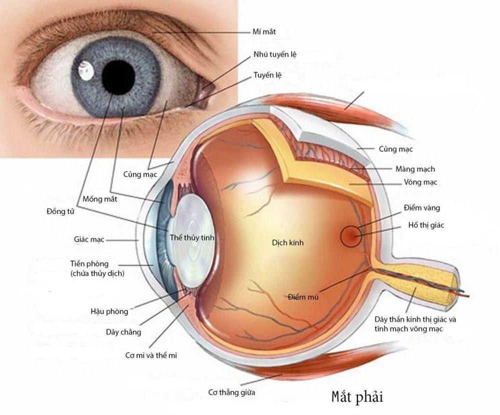
The eyeball is spherical, located in a bone cavity (orbital). Orbital anatomy is relatively complex. The axial length of the eyeball in an adult is about 22-24mm. Short or long eyeball causes refractive errors such as nearsightedness or farsightedness.
Structure of the eye in the ophthalmic region includes the following parts:
1.1 Sheath of the eyeball The sheath of the eyeball includes:
The cornea: is a transparent membrane, very tough, without blood vessels, has a spherical cap, occupies Anterior 1/5 of the eyeball. The cornea is about 11mm in diameter, the radius of curvature is 7.7mm, the thickness at the edge is 1mm, in the center is 0.5mm, the refractive power is about 45D. The cornea has layers from the outside to the inside, including: epithelium, Bowman's membrane, parenchyma, and endothelium. The cornea is nourished by tears, aqueous humor and osmosis from the periphery of the blood vessels; Sclera: A very tough, white fibrous tissue that occupies 4/5 of the back of the eyeball. The sclera is made up of many thick, criss-crossed fibrous bandages that protect the membranes and internal environments. The thickness of the sclera depends on the region: thickest in the posterior pole (1 - 1.35mm), thinnest at the attachment of the axial muscles (0.3mm). The posterior sclera pole has a hole with a diameter of 1.5mm, covering the hole is a sclera with many small holes that allow the optic nerve fibers to pass. 1.2 Choroid The choroid (uvea) consists of 3 parts: iris, ciliary body and choroid. The iris + ciliary body is called the anterior uvea, the choroid is called the posterior uvea. In eye anatomy, the uvea is responsible for nourishing the eyeball and regulating eye pressure.
The uvea includes:
Iris: Has a hole in the center of the coin, the front is the posterior limit of the anterior chamber, blue, black or brown depending on the race. The posterior surface of the iris is uniformly dark brown, which is the anterior limit of the posterior chamber. There is a circular hole in the middle of the iris (pupil). Histologically, the iris consists of 3 main layers: the endothelial layer on the anterior surface, the stroma, and the epithelial layer on the posterior surface. The main role of the iris is to regulate the amount of light reaching the retina (through changing the pupil size); The ciliary body: The raised part of the uvea, located between the iris and the choroid. The ciliary body has a regulatory role in helping the eyes see clearly near objects, secreting aqueous humor (thanks to cells in the fascia). The ciliary body hidden behind the iris is an irregular circular band. Seen from the back, the ciliary body has 2 parts: the posterior part (the arc of the ciliary body) and the anterior part (the ciliary body). In terms of histology, from the outside to the inside of the ciliary body, there are layers: the upper ciliary layer, the ciliary body layer, the vascular layer, the vitreous layer, the pigment epithelium, the ciliary epithelium and the inner limit layer; Choroid: A loose connective tissue located between the sclera and the retina. The choroid has many blood vessels and black pigment cells, which are responsible for feeding the eyeball, turning the lumen of the eyeball into a dark chamber, helping the image to be displayed clearly on the retina. In terms of organization, from outside to inside the choroid consists of 3 layers: true choroidal layer, genuine choroidal layer, Bruch's membrane. Blood vessels and nerves of the uvea according to eye anatomy include:
Arteries: The uvea has 2 systems: the posterior short ciliary artery (consisting of about 20 arteries) and the posterior long ciliary artery (with 2 arteries). circuit); Veins: Blood from the uvea follows small veins, gathers in 4 large veins, goes out of the eyeball, flows through the eye veins into the cavernous sinuses; Lymph: Uvea is absent; Nerve: Consists of long ciliary nerve fibers and short ciliary nerve fibers.

Cấu tạo mắt với phần nhãn cầu có hình cầu trong ổ mắt
1.3 Retina The retina, also known as the neural membrane, is located in the lumen of the uvea. This is the place to receive light stimuli from the outside, transmitted to the visual analysis center in the cerebral cortex.
In terms of shape, the retina consists of 2 parts: the sensory retina and the insensitive retina, located about 7-8mm from the edge of the cornea. The center of the retina (corresponding to the posterior pole of the eyeball) is a light-colored area (macular). In the middle of the macula, there is a small concave pit (central pit). 3.4 - 4mm away from the macula to the nose is the optic nerve - the starting point of the optic nerve. Optic spines are round or oval, about 1.5mm in diameter, pale pink, clearly demarcated with surrounding areas. In terms of structure, the retina has 4 layers of cells: the pigment epithelial layer, the visual layer, the bipolar cell layer, and the ganglion cell layer (multipolar cells).
Blood vessels of the retina:
Central retinal artery: Is a branch originating from the ophthalmic artery, running to the eyeball, about 10mm from the posterior pole of the eyeball, then it enters the optic nerve and goes to the retina; Veins: Usually parallel to arteries. 1.4 Front room and back room When doing eye surgery, it is impossible not to mention the front room and the back room.
Anterior chamber: Is a cavity located between the cornea in front, iris and vitreous behind, filled with fluid. The central part of the anterior chamber is the deepest (about 3 - 3.5mm), the closer to the edge, the deeper the depth decreases. The anterior chamber angle at the lateral margin is bounded by the cornea - sclera anteriorly, iris - ciliary body posteriorly. The anterior chamber has an important physiological and surgical role. The anterior chamber has the following components: ring of Schwalbe, Trabeculum, canal of Schlemm, sclera spur, ciliary body strip, iris root; Posterior chamber: The posterior chamber has an anterior limit of the posterior surface of the iris, and the posterior limit of the anterior surface of the vitreous membrane. The back room is connected to the front room through the pupil hole. Inside the back room contains fluid like the front room. 1.5 Transparent media The transparent media inside the eyeball include:
Aqueous: A transparent liquid secreted by the ciliary body, filled inside the anterior and posterior chambers. The composition of aqueous humor includes water, albumin, globulin, glucose, amino acids,... The aqueous humor plays the role of a factor affecting intraocular pressure, providing nutrition for the vitreous, contributing to corneal nourishment; The vitreous body is a transparent, biconvex lens that is fixed to the ciliary body by Zinn cords. The thickness of the ciliary body is about 4mm, the diameter is 8-10mm, the radius of curvature is 10mm at the front, the back is 6mm, the optical power is about 20-22D. The crystalline lens has two faces, anterior and posterior, which meet at the equator. The vitreous body consists of three parts: the capsule, the subcapsular epithelium, and the fibers of the vitreous. The vitreous has no blood vessels or nerves and is nourished by selective osmosis from aqueous humor. The vitreous body plays an important role in the refractive system, helping to focus on the correct focus of the image on the retina when looking at a distance, helping the eyes to see clearly near objects (thanks to accommodation); Vitreous: Is a liquid like egg white, located behind the vitreous, occupying the entire posterior part of the eyeball. The main component of vitreous is a fibrous structural protein (Vitrein), with hyaluronic acid filling in the spaces between the fibers.
2. Eye Anatomy: Eyeball Protectors
2.1 Eye sockets There are 2 eye sockets located on either side of the nasal cavity, made up of the skull and facial bones. The eye socket has a pyramidal shape, 4 sides with 4 bone walls, the apex turns to the back, the base turns to the front.
Characteristics of the orbit:
Size: In an adult, the volume of the orbit is about 29ml, the height from the top to the bottom of the orbit is about 40mm, the width of the base of the orbit is about 40mm, the height of the fundus is about 35mm ; The walls of the orbit: Upper wall (orbital ceiling), outer wall, lower wall (orbital base), medial wall; The base of the eye socket: It is oval in shape, consisting of 4 banks (upper, outer, lower and inner); Top of eye socket: With visual hole and 1 V-shaped slit; Elements located in the orbit: Ocular motor muscles (6 muscles including 4 rectus and 2 oblique muscles), muscles of the eyelids (upper eyelid muscle, sphincter muscle), intraorbital tendons (fibrous membrane) periorbital muscle, Tenon capsule), organization of the fovea; Eyelids: Each eye has 2 eyelids (upper and lower eyelids), anatomically similar. The eyelids have 4 layers from front to back, including: eyelid skin, eyelid muscle layer (eyelid sphincter, upper eyelid lifter muscle), eyelid cartilage layer, and conjunctival layer. The ciliary circulation includes arteries and veins. 2.2 Tears In the anatomy of the eye, the lacrimal gland consists of two parts:
Tear secretory: Tears are secreted from the main lacrimal gland (located in the upper outer corner of the orbit) and the accessory lacrimal gland (scattered in the eye) conjunctiva). Tears are responsible for providing nutrition and protecting the cornea; Tear path: Surface water is collected into the upper and lower lacrimal holes, enters the upper and lower lacrimal glands, passes through the common lacrimal duct, and collects into the lacrimal sac. From here, tears pass through the lacrimal duct, down the nose at the lower nasal passage.

Cấu tạo mắt với vị trí của lệ bộ và hốc mắt
3. Eye anatomy: Nerve line and visual mid-autumn
3.1 Optic Nerve The axon of ganglion cells will focus on the optic nerve, pass through the ethmoid, and form the optic nerve (second nerve). The optic nerve travels to the top of the fovea and passes through the optic foramen into the skull. Then, axons of ganglion cells of the nasal hemiretinal (nasal bundle). Cross to the opposite side to go with the other temporal bundle, stopping at the lateral knee. The position where the two nasal bundles cross each other is called the visual interference (located above the pituitary).
The segment from the junction to the lateral commissure has fibers that tend to spread out, so it is called the visual band. From the lateral kneecap, the visual fibers continue to expand, so called optic rays, to stop at the cortex of the occipital lobe.
Circulation of the optic nerve is ensured by three arteries: the anterior choroidal artery, the posterior cerebral artery and the Sylvius artery.
Includes cortical regions 17, 18, 19 belonging to the cerebral cortex and occipital lobes, around the striatum partially encroaching on the outer surface of the occipital lobe.
Eyes are one of the most important organs in the body. Understanding the anatomy of the eye and the function of the parts of the eye will help each person be more conscious in protecting the health of their eyes.
Follow Vinmec International General Hospital website to get more health, nutrition and beauty information to protect the health of yourself and your loved ones in your family.
Please dial HOTLINE for more information or register for an appointment HERE. Download MyVinmec app to make appointments faster and to manage your bookings easily.


