This is an automatically translated article.
Acute cor pulmonale includes all cases of acute right heart failure due to a sudden increase in acute pulmonary vascular resistance. Pulmonary embolism is an occlusion of at least one pulmonary artery or pulmonary artery, usually due to ascending deep vein thrombosis. This is a fairly common disease, but difficult to diagnose because it is easily confused with other diseases. The following article clarifies the symptoms, diagnosis and management of acute bronchiolitis and pulmonary embolism.
1. Symptoms, how to diagnose acute bronchiectasis 1.1. Onset symptoms Patients with acute bronchiectasis often have the same symptoms as:
Sudden feeling of sharp pain in the chest, unable to breathe as usual. Presternal angina pectoris appeared in varying degrees due to functional coronary insufficiency. Angina radiates to the shoulders, arms, and sometimes to the abdomen and examines the patient for a reaction to the abdominal wall. The patient has difficulty breathing rapidly and shallowly, coughs up pink foam. There is a feeling of dread every time the pain occurs. After a brief hypertensive crisis, the systolic blood pressure will drop, the blood pressure may be stuck, and in severe cases. At that time, the patient may fall into a state of collapse, cyanosis and shock. This condition occurs very early and the right heart failure is more severe the bigger the blockage.
1.2. Diagnosis of acute bronchiectasis 1.2.1. Clinical examination Tachycardia is an invariable symptom, usually sinus tachycardia, but transient complete arrhythmias are not uncommon.
The doctor can examine the Harzer sign, hear the heart with a galloping sound of the right ventricle. The second heart sound heard in the pulmonic fossa is intense or may be sharp. In addition, a transient pericardial rub may be heard. The systolic murmur due to functional tricuspid regurgitation is no exception.
Peripheral signs of right heart are also common: jugular venous distension, sometimes palpitations. Enlarged and painful liver, positive jugular hepatobiliary reflex, sometimes more rarely, the doctor will feel the area of the liver beating with the heartbeat. Central venous pressure is usually elevated above 20cm of water.
Lungs and pleura generally show no specific signs. But on examination, rales can be heard in the lungs, scattered or localized, reflecting pulmonary edema.
It is very important to look for and detect signs of peripheral thrombophlebitis, especially in the lower extremities. The signs of thrombophlebitis are generally very subtle, requiring a very meticulous examination to be detected: the area of thrombophlebitis has a higher than normal temperature, localized warmth to the touch, the veins have a warm sensation to the touch. Dilated superficial pulses, decreased swinging ability of the lower leg, anterior ankle edema, pain when flexing the foot sharply into the lower leg (Homans sign).
Usually it is recognized that thrombophlebitis manifests completely latent and silent, with no focal signs, and its detection when performing tests such as exploration of the venous system of the lower extremities by Doppler in these patients.
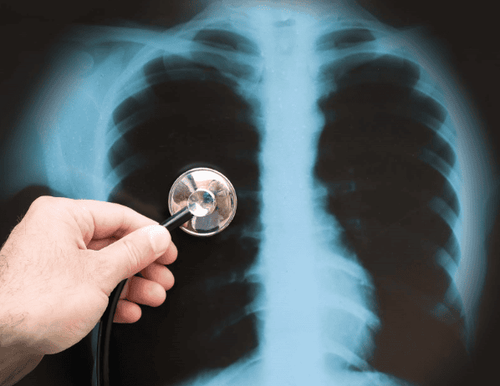
Chụp X – quang chẩn đoán tâm phế cấp
1.2.2. Chest X-ray During the first 2 or 3 days, the chest X-ray is usually nothing special. However, the doctor can see dilated right atrial arch, pulmonary artery distension.
In the case of a large occlusion one can see the pulmonary artery branch dilated then the lower segment shrink or suddenly become indistinct as if amputated.
Some other cases can see too bright image of the lung parenchyma showing signs of no circulation in this area. Occasionally, radiographs show an uneven appearance due to pulmonary edema.
1.2.3. Electrocardiogram Sinus tachycardia is a common sign. However, atrial fibrillation or atrial flutter is not uncommon, especially when pulmonary embolism affects the heart.
Signs of right atrial hypertrophy (Pneumothorax) are in fact not many. In contrast, variable ventricular complexes are quite diverse.
In the peripheral leads, the heart rotates clockwise creating the S1Q3 image that has been described by MarGin and White, the Q3 waves are deep but thin, not dark, in contrast to the portal Q3 wave. necrotic heart muscle. In principle, there should be no Q waves in aVF but this rule really has many exceptions. This image of S1Q3 will be even more valuable if it was not available on previous electrocardiograms. The normal telegraph axis shifts to the right. T waves are usually found to be negative at D3, positive or biphasic at D2. In the precordial leads, subendocardial ischemia in the anterior septal region is a common finding, presenting with symmetric, sharp, negative T waves in V2, V3, and aVF. It may initially be accompanied by a St-segment gradient indicating transient ischemia. This sign generally disappears after several weeks. The presence of a constant positive T wave in lead V1 is the main feature distinguishing it from anterior septal myocardial infarction. Incomplete right bundle branch block is more common because this ECG sign is very early and transient. More rarely, complete right bundle branch block, however, represents a very poor prognosis for the patient. The above-mentioned combination of multiple abnormal electrocardiographic findings is characteristic of pulmonary embolism. When they appear individually, evaluation becomes more conservative. For proper analysis, it is necessary to compare multiple ECGs in different intervals to draw conclusions. 1.3. Progression and prognosis Immediate progression of acute bronchiectasis is generally difficult to predict. However, the presence of acute bronchiectasis significantly worsens the prognosis of pulmonary embolism.
Sometimes suddenly the disease progresses in a very bad direction. The patient entered a state of severe cardiovascular collapse and shock, severe cyanosis, severe respiratory failure, marked right-sided heart failure, complete right bundle branch block, signs of widespread myocardial ischemia. Advanced age is a poor prognostic factor.
In well-progressed cases, chest pain, shortness of breath, and expression of fear will gradually disappear. However, these symptoms will persist and improve after 24 to 36 hours. Patients may be accompanied by symptoms: expectoration of rust-colored, thick, garlic-smelling sputum or just coughing up mucus and blood. The temperature rose from 38 to 38.5 degrees on day 2. Cardiovascular signs gradually decreased. The lungs may form infarcts, as indicated by pulmonary consolidation or broncho-alveolar inflammation.
Appearance of pleural fluid due to pleural reaction is a very common symptom. In general, the number of translations is usually small. The discharge is lemon yellow or pink, with a lot of albumin, positive Rivalta reaction.
Later progress is usually favorable. Pulmonary infarction will disappear after a few weeks. Superinfection or infarct abscess is uncommon. Some cases of pulmonary infarction recur many times over a long period of time despite effective anticoagulation. These cases lead to a very poor prognosis, that is, cor pulmonale after pulmonary infarction.
2. Symptoms, how to diagnose pulmonary embolism (pulmonary embolism) 2.1. Symptoms of pulmonary embolism Manifestations of pulmonary embolism can vary from sudden hemodynamic collapse to progressively progressive dyspnea. (The patient's previous poor cardiopulmonary status is an important predisposing factor to hemodynamic collapse.) Most patients with pulmonary embolism are asymptomatic at presentation. In contrast, patients with symptomatic DVT often have pulmonary embolism confirmed on diagnostic studies in the absence of pulmonary symptoms. Sickle cell disease often makes diagnosis difficult in relation to pulmonary embolism. A chest infection is often the presenting symptom.
Patients with pulmonary embolism may have atypical symptoms. In such cases, a clear suspicion of pulmonary embolism based on the presence of risk factors may lead to consideration of pulmonary embolism in the differential diagnosis. These symptoms include:
Convulsions Syncope Abdominal pain Tachycardia (> 100 beats/min) Fever above 37.8 degrees Cough with phlegm Wheezing Lower extremities Decreased level of consciousness New onset of atrial fibrillation Rib pain Delirium (in elderly patients) 2.2. Diagnosis The initial assessment of pulmonary embolism requires pulse oximetry and chest radiographs. Electrocardiogram, arterial blood gas (ABG), or both may help rule out other diagnoses (eg, acute myocardial infarction).
2.2.1. Chest radiograph This technique is usually nonspecific but may show atelectasis, focal infiltrates, an elevated hemidiaphragm, or pleural effusion. The classical findings of focal loss of pulse markers (Westermark sign), peripheral density, wedge-shaped pleural origin (Hampton's tumor), or enlargement of the right descending pulmonary artery are suggestive but uncommon (i.e. insensitive) and low in specificity. This method can also help rule out pneumonia. Pulmonary infarction due to pulmonary embolism can be confused with pneumonia.
2.2.2. Oximeter technique This method provides a quick way to assess oxidation; Hypoxemia is a sign of PE and it requires further evaluation. Blood gas testing should be particularly considered for patients with dyspnea or tachypnea who are not hypoxic detected by pulse oximetry.
Measurement of arterial or venous blood gases may show an increased alveolar oxygen difference with arterial oxygen (Aa) increased (sometimes referred to as the Aa gradient) or decreased blood CO2. Both of these tests are moderately sensitive for PE, but not specific. Blood gas testing should be particularly considered for patients with dyspnea or tachypnea who are not hypoxic detected by pulse oximetry. Oxygen saturation may be normal due to a small clot burden, or due to compensatory hyperventilation; Very low pCO2 detected by ABG measurement can confirm hyperventilation.
2.2.3. This technique often shows tachycardia and various ST-T wave abnormalities, which are not specific for pulmonary occlusion. S1Q3T3 or a new right bundle branch block may show the effect of a sudden increase in RV size affecting the RV pathways; These findings have moderate specificity but are not sensitive, occurring in only about 5% of patients, although these findings occur in a higher proportion in patients with severe PE. There may be right axis deviation (R > S in V1) and P-pulmonale. T wave inversion in leads V1 to V4 also occurs.

Biểu hiện của tắc động mạch phổi có thể thay đổi từ suy sụp huyết động đột ngột đến khó thở tiến triển dần dần
2.2.4. CT angiography This is the preferred imaging technique for the diagnosis of acute pulmonary embolism. CT angiography results are fast, accurate, and highly sensitive and specific. This method can also provide additional information about other lung disease (eg, demonstrating pneumonia rather than PE as a cause of hypoxia or pleuritic chest pain) as well as the severity of the disease. severity of PE (eg, size of the right ventricle or regurgitation into the hepatic vein).
Although poor quality scans due to motion artifacts or poor contrast can limit the sensitivity of the examination. But improvements in CT technology have shortened acquisition time to less than 2 seconds, providing relatively motion-free images in patients with dyspnea. Fast scan times allow smaller amounts of iodinated contrast to be used, which reduces the risk of acute kidney injury.
Sensitivity of CT angiography is highest for pulmonary artery occlusion in the main pulmonary artery and in the lobar and segmental vessels. The sensitivity of CT angiography is lowest for branch vessel embolism (about 30% of total pulmonary embolism). However, the sensitivity and specificity of CT angiography has improved as the technology has progressed.
2.2.5. Ventilation/perfusion (V/Q) tomography (V/Q) Ventilation/perfusion (V/Q) tomography in pulmonary embolism helps to detect areas of the lung that are ventilated but not perfused. V/Q scans take longer than CT angiograms and are less specific.
However, when chest X-ray results are normal or near-normal and there is no significant underlying lung disease, it is a highly sensitive test. V/Q imaging is particularly useful when renal failure precludes the use of another required contrast agent for CT angiography.
In some hospitals, the V/Q scan can be done with a portable machine that provides 3 views of ventilation and perfusion, which is useful when the patient is too sick to move. Perfusion defects can occur in many other lung conditions (eg, COPD, pulmonary fibrosis, pneumonia, pleural effusion). Inappropriate perfusion defects that can mimic PE can occur in pulmonary vasculitis, pulmonary venous thrombosis, and sarcoidosis.
2.2.6. Duplex Ultrasound This method is a safe, non-invasive, portable technique for detecting blood clots in the leg or arm. A blood clot can be detected by showing poor venous compression or by showing reduced flow using Doppler ultrasound.
The test has > 95% sensitivity and > 95% specificity for thrombosis. Confirmation of a DVT in the calf or iliac vein can be more difficult but is generally achievable. The sonographer should always attempt to image the underlying popliteal vein into its triangular portion.
The absence of thrombosis in the leg veins does not rule out the possibility of thrombosis from other sources, such as the upper extremity, but patients with suspected DVT and negative results on duplex Doppler have > 95% survival Uncomplicated survival, as thrombosis from other sources is much less common.
2.2.7. Echocardiography Echocardiography may reveal a blood clot in the right atrium or ventricle, but echocardiography is most commonly used for risk stratification in acute PE. The presence of right ventricular dilatation and reduced mobility may indicate the need for more aggressive treatment.
2.2.8. Pulmonary angiography Pulmonary angiography is now rarely needed to diagnose acute PE because noninvasive CT angiography has similar sensitivity and specificity. However, in patients receiving catheter-based thrombolytic therapy, pulmonary angiography is useful to assess catheter placement and can be used as a rapid means of determining the success of a catheter. procedure when the catheter is removed.
Pulmonary angiography is also still used along with right-heart catheterization to assess whether a patient with chronic pulmonary arterial hypertension is a candidate for endocarditis.
2.3. Prognosis An estimated 10% of patients with pulmonary embolism die within the first few hours after presentation. Most patients who die from acute PE are never diagnosed before death. In fact, PE is not suspected in most of these patients. The best prospects for reducing mortality are related to
Improve diagnostic frequency (eg, by including PE in the differential diagnosis when patients have nonspecific but compatible symptoms or signs or symptoms. ) Improves promptness of diagnosis Improves risk stratification Improves promptness to initiate anticoagulation Provide appropriate prophylaxis in at-risk patients Very high D-dimer levels High seems to predict a poor outcome.
Patients with chronic thromboembolic disease represent a small, but important portion of PE survivors. Anticoagulation treatment reduces PE recurrence rates by about 5%, and some studies have found anticoagulants reduce recurrence rates even lower.

Có thể sử dụng tất cả các thuốc an thần làm dịu cơn đau
2.4. Treatment 2.4.1. Anti-pain and sedation All sedative drugs can be used to relieve pain, fear and anxiety for patients except Morphine because the drug can cause respiratory depression.
2.4.2. Ensure ventilation. Give oxygen through a tube from 6 to 8 liters/min.
2.4.3. Anticoagulation Treatment Initial anticoagulation options for acute PE include IV unfractionated heparin Low molecular weight heparin subcutaneously Ficaparinux subcutaneous Factor Xa inhibitors (apixaban and rivaroxaban) Argatroban injection Intravenous administration of heparin-induced thrombocytopenia Intravenous unfractionated heparin has a short half-life (useful when the potential for bleeding is higher than normal) and is reversible with protamine administration. An initial bolus of unfractionated heparin was given, followed by a blend of heparin doses by protocol to achieve an activated PTT 1.5-2.5 times that of normal control. Therefore, non-fractionated heparin requires ongoing hospitalization for management. Furthermore, the pharmacokinetics of unfractionated heparin are relatively unpredictable, leading to frequent episodes of over- and under-anticoagulants and the need for frequent dose adjustments. Despite this, many clinicians prefer this unfractionated heparin regimen, particularly when thrombolytic therapy is given or contemplated or when the patient is at risk of bleeding because if bleeding occurs, The short half-life means that the anticoagulant is rapidly reversed once the infusion is stopped.
In patients with renal impairment, dose reduction is required, and subsequent verification of appropriate dosage should be done by testing serum factor Xa levels (target: 0.5 to 1.2 IU/ mL measured 3 to 4 hours after the 4th dose). Low molecular weight heparin is generally contraindicated in patients with severe renal impairment (creatinine clearance <30 mL/min). Low molecular weight heparin is partially reversible with protamine.
Fondaparinux is a factor Xa antagonist that is injected subcutaneously. It can be used in acute DVT and acute PE instead of heparin or low molecular weight heparin. It has also been shown to prevent recurrence in patients with superficial venous thrombosis. Results appear to be similar to total heparin heparin . The advantage is that a fixed dose is administered once or twice a day, without the need for monitoring of anticoagulation levels, and with a reduced risk of thrombocytopenia. The dose (in mg/kg x 1 time/day) is 5 mg for patients < 50 kg, 7.5 mg for patients 50-100 kg, and 10 mg for patients > 100 kg. Fondaparinux dose reduction by 50% if creatinine clearance is between 30 and 50 mL/min (0.5 - 0.83 mL/s). The drug is contraindicated if the creatinine clearance is <30 mL/min.
2.4.4. Maintenance anticoagulation Maintenance anticoagulation is indicated to reduce the risk of clot expansion or embolism and to reduce the risk of new clot formation. Drug options for maintenance anticoagulation include
Oral vitamin K antagonists (warfarin in the US) Oral factor Xa inhibitors (apixaban, rivaroxaban, edoxaban) Oral direct thrombin inhibitors (dabigatran) Rare when administering low molecular weight heparin subcutaneously Warfarin is an effective long-term oral anticoagulant option that has been used for decades, but it is inconvenient for a number of reasons. In most patients, warfarin is started on the same day as the heparin (or fondaparinux) therapy used for initial anticoagulation. Heparin (or fondaparinux) therapy should be combined with warfarin therapy for a minimum of 5 days and until the INR is within the therapeutic range (2.0 to 3.0) for at least 24 hours.
The main disadvantages of warfarin are the need for periodic INR monitoring, frequent dose adjustments, and drug interactions. Physicians prescribing warfarin should be alert for drug interactions; In a patient taking warfarin, almost any new drug should be tested.
Bleeding is the most common complication of warfarin treatment; patients > 65 years of age and those with comorbidities (especially diabetes, recent myocardial infarction, hematocrit < 30%, or creatinine > 1.5 mg/dL [ > 133 micromol/L]) and A history of stroke or gastrointestinal bleeding appears to be at greatest risk. Bleeding can be reversed with vitamin K 2.5 to 10 mg IV or orally and, in an emergency, with fresh frozen plasma or a new concentrate (prothrombin complex concentrate) containing factor II (prothrombin), factor VII, factor IX, factor X, protein C, and protein S. Vitamin K can cause flushing, local pain and, rarely, anaphylaxis.
2.4.5. Treatment with muscle-enhancing agents Cardiogenic shock occurs in more than 50% of cases of pulmonary embolism. Treatments include ventilatory support, if hypoxia is persistent and prolonged, drugs that increase myocardial contractility, thrombolytic therapy, or surgical removal of the clot in the event of failure or resistance. indications for the use of thrombolytic agents. Treatment with inotropic agents will depend on cardiac output and pulmonary arterial pressure as measured by a Swan-Ganza catheter.
The doctor will prescribe the use of a continuous intravenous infusion pump with a dose of 2.5 - 15 mcg/kg/min. Adrenaline (0.01–0.05 mcg/kg/min) or Dopamine at doses between 2.5 and 15 mcg/kg/min can also increase cardiac output by potentiating myocardial and vasodilating effects.
2.4.6. Systemic thrombolysis Systemic thrombolytic therapy with alteplase (tissue plasminogen activator [tPA]) provides a non-invasive method to rapidly restore pulmonary blood flow, but its use also causes Arguably the long-term benefit does not outweigh the obvious bleeding risk. Nevertheless, most experts agree that systemic thrombolytic therapy should be used in hemodynamically compromised patients, especially when the disease is severe. Although no single prospective randomized trial of systemic thrombolytic therapy has shown improved survival in patients with subsevere PE, some experts recommend thrombolysis. mass, especially when the patient also has multiple or large clots, very severe RV dysfunction, marked tachycardia, significant hypoxemia, and other concomitant findings such as residual clot legs, positive troponin values and/or elevated BNP values. Others reserve thrombolytic therapy only for patients with severe (high-risk) PE. Streptokinase and urokinase are generally no longer used.
Absolute contraindications to thrombolytics include
Before hemorrhagic stroke Ischemic stroke within 1 year Active external or internal bleeding from any source Intracranial trauma or surgery within 2 months Intracranial tumor Certain surgeries within the previous few days Relative contraindications include
Recent surgery (≤ 10 days) Visceral bleeding (as in liver failure) Pregnancy recent perforation of large incompressible veins (eg, subcutaneous or internal jugular veins) Recent femoral artery catheterization (eg, 10 days) Peptic ulcer disease or other other conditions that increase the risk of bleeding Severe hypertension (systolic blood pressure > 180 mm Hg or diastolic blood pressure > 110 mm Hg) Head trauma from PE due to syncope, even with a normal brain CT 2.4.7 . Surgical treatment Only reserved for PE patients with hypotension despite supportive measures (persistent systolic blood pressure 90 mmHg after fluid and oxygen therapy or if vasopressor therapy is required). pulse) or is on the verge of cardiac arrest or respiratory arrest. Surgical resection should be considered if thrombolysis is contraindicated; In such cases, catheter-directed vortex resection may also be considered and, depending on local resources and expertise, attempted before embossing. Surgical excision of the throat appears to increase survival in patients with severe PE but is not widely available.
Please dial HOTLINE for more information or register for an appointment HERE. Download MyVinmec app to make appointments faster and to manage your bookings easily.