This is an automatically translated article.
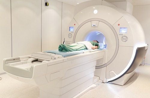
The article was professionally consulted with Specialist Doctor I Nguyen Dinh Hung - Diagnostic Imaging - Department of Diagnostic Imaging - Vinmec Hai Phong International General Hospital.The abdominal cavity contains many complex anatomical structures of the cardiovascular, digestive and urinary systems. Therefore, when there is an injury of an intra-abdominal organ, it will be very complicated and difficult to evaluate. Abdominal magnetic resonance imaging is a very valuable means of detecting, assessing the extent of injury and monitoring abdominal pathology. So what is the procedure for magnetic resonance imaging of the abdomen without injecting magnetic contrast?
1. Outline
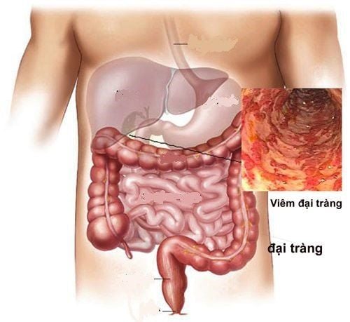
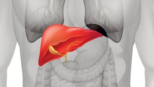
The organs of the digestive system located in the abdomen include the stomach, duodenum, small intestine, colon, liver, gallbladder, and pancreas. In addition, the urinary system is also located in the abdomen including the kidneys and urinary tract. The spleen, adrenal glands, and major blood vessels are also essential anatomical structures of the body.Magnetic resonance imaging (MRI) of the abdomen is an essential imaging method in patients with suspected abdominal injuries because the images from MRI have good contrast, high resolution, and clear images. It helps the doctor to easily assess the injury. Moreover, magnetic resonance imaging (MRI) also has the ability to reproduce 3D images, and visualize anatomical structures that need to be evaluated to help improve the ability to diagnose pathology.
Abdominal magnetic resonance imaging (MRI) is an increasingly improved and updated technique involving multiple pulse sequences. The shooting process is constantly changing and improving. In this article, we will refer to the abdominal magnetic resonance imaging procedure without injection of magnetic contrast (excluding the liver).
2. Indications for abdominal magnetic resonance imaging without injection of contrast agent
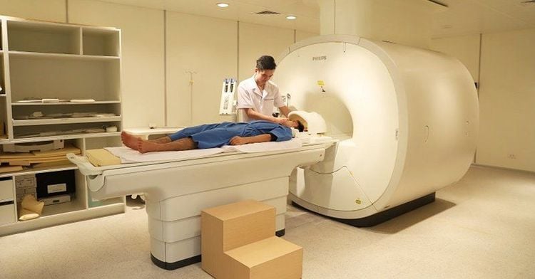
Indications for abdominal magnetic resonance imaging are usually indicated after having images of suspected lesions from imaging tools such as ultrasound, X-ray. Then, depending on the disease at the organ, the doctor will give appropriate indications.● Pancreatic diseases such as: pancreatic tumor, evaluate for suspected pancreatic lesions or unexplained proliferation, evaluate for obstruction or dilatation of pancreatic duct or pancreatic juice leak, pancreatic duct obstruction, pancreatic juice leak. Evaluation of chronic pancreatitis, complicated acute pancreatitis. Preoperative evaluation of pancreatic tumors. Follow-up after pancreatic surgery.
● Diseases of the spleen such as detecting and diagnosing suspected lesions in the spleen after initial diagnostic methods, assessment of diffuse splenic lesions, assessment of suspected subsplenic tissue.
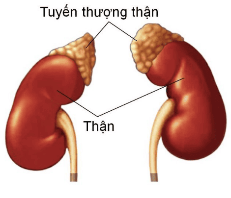
● Urinary system pathologies: Surveying the kidneys, ureters and retroperitoneal tumors, detecting renal tumors, preoperative assessment of renal tumors including assessment of the renal vein and inferior vena cava. Evaluation of the urinary tract for anatomical abnormalities (MR urography). Follow-up after surgery, renal tumor intervention.
● Diseases of the adrenal glands: adrenal tumors (functional tumors and pheochromocytoma).
Digestive system diseases: detecting and monitoring after treatment of cholangiocarcinoma, gallbladder. Detection of gallstones or biliary stones, degree of biliary dilatation, congenital malformations of the biliary tree. Tumors in the stomach, rectum, small intestine, mesentery.
3. Contraindications to magnetic resonance imaging without contrast injection
Absolute contraindications: carrying electronic devices with metal, metal surgical instruments < 6 months, patients in emergency - intensive resuscitation. Relative contraindications: metal surgical facilities > 6 months, phobia of the dark or solitude.4. Preparation steps for magnetic resonance imaging without contrast injection
Medical staff: radiologist and radiologist.Resonance machine 1.5 Tesla or more
● Medicine: sedation, oral contrast agent or enema.
● Patients: need to be explained the procedure, place an intravenous line, depending on the purpose of the survey, there will be oral contrast or enema through the anus.
5. Steps to conduct abdominal magnetic resonance imaging without injecting contrast material
5.1. Some note
Use the transducer coil in the face, except in cases where the patient has a medical condition that cannot be placed. Select the field of view (FOV) with the highest resolution and best signal/noise ratio. Most cases of abdominal evaluation use the T1W and T2W imaging sequences. Acquiring images in many different planes, the thickness of the cut layer does not exceed 1 cm with the interval between the layers not exceeding 3 mm.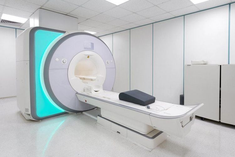
5.2. Selective pulse sequences in non-contrast MR imaging of the abdomen
● To generate T1W images, conventional spin echo pulse sequences, echo train spin echo (TSE) or fast spin echo (FSE) or gradient (echo) are used.● T2W imaging can be done using fast spin echo or hybrid gradient and spin echo (GRASE) techniques. Defatting can take the form of inversion recovery (STIR), selective chemical fat saturation or spectral presaturation inversion recovery (SPIR), or Dixon techniques and water stimulation.
● Three-dimensional (3D) techniques can produce both T1W and T2W images providing high resolution, higher signal-to-noise ratio.
● Use of magnetic contrast agents is beneficial in the gastro-intestinal survey. When used orally to examine the small intestine, the lumen-forming agent is black on T1-weighted images, helping to better detect lesions in the intestinal wall.
● In-phase and out-phase pulse sequences help detect intracellular lipids in adrenal tumors (adenomas), kidneys (clear cell carcinomas) and to confirm fatty infiltration of organs such as the pancreas.
● T2W series of cholangiopancreatography (MRCP) helps to examine the morphology and anatomy of the biliary tract and pancreatic duct. Create a T2W image using the acquisition relaxation enhance (RACE) pulse sequence. In addition, the use of secretin helps to increase the visibility of the pancreatic duct to help diagnose anatomical changes, chronic pancreatitis, intraductal mucinous-papillary tumors, and quantify exocrine pancreatic function.
6. Read the results
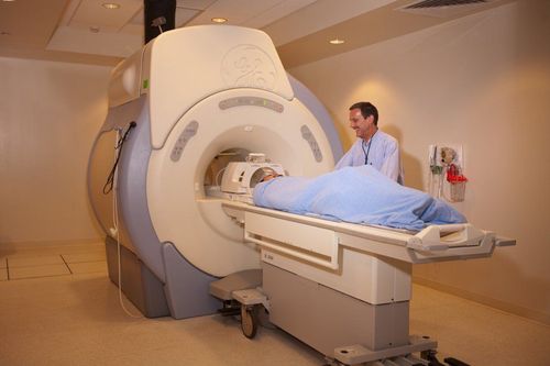
Results are printed to a movie or saved to a CD. The doctor reads the lesions described on the machine and can give professional advice to the patient and family if necessary. Especially, Vinmec International General Hospital is the first unit in Southeast Asia to put into use the scanner. Benefit from the new 3.0 Tesla Silent technology from the US manufacturer GE Healthcare. The 1.5 tesla magnetic resonance imaging machine has many outstanding advantages such as:Non-invasive and highly safe. Magnetic resonance imaging with 1.5 tesla MRI machine is not exposed to X-rays. There are no contraindications for pregnant women and young children, expanding the scope of diagnosis and treatment. Provides high resolution images that evaluate the structure of organs and soft tissues in detail. Detects small-sized lesions, in difficult-to-identify locations. Shorten the imaging time compared to low tesla magnetic resonance machines. The noise when operating the 1.5 tesla MRI machine has been much reduced compared to the older generations, Contrast, if used when taking the 1.5 tesla MRI machine, is less likely to cause side effects such as anaphylaxis . The machine currently applies the safest and most accurate magnetic resonance imaging technology available today, without using X-rays, non-invasively. Silent technology is very beneficial for patients who are young children, the elderly, patients with weak health or have just had surgery.
Currently, Vinmec International General Hospital with the motto: Always put the patient at the center, Vinmec is committed to bringing comprehensive and high-quality healthcare services to customers.
With modern facilities of international standards, quality medical services, a team of medical professionals, experienced doctors will bring optimal treatment results to customers.
Friendly hospital environment, international standard pre-, post-operative care facilities will bring the most accurate diagnosis results, best treatment, fastest health recovery.
Please dial HOTLINE for more information or register for an appointment HERE. Download MyVinmec app to make appointments faster and to manage your bookings easily.


