This is an automatically translated article.
The article is professionally consulted by Dr. Nguyen Dinh Hung - Diagnostic Imaging - Department of Diagnostic Imaging - Vinmec Hai Phong International General Hospital.Magnetic resonance imaging (MRI) is a common imaging method worldwide. Doctors and researchers are constantly improving MRI techniques to aid in the accurate diagnosis of patients. This article looks at the 6 most basic things about MRI scans you need to know.
1. Reasons for doing an MRI
Magnetic resonance imaging: is one of the most popular imaging methods in the world, especially for soft tissues and nervous system. There are many reasons for doctors to prescribe MRI scans such as:
Magnetic resonance imaging of the brain and spinal cord to determine:
● Damage to brain blood vessels;
● Traumatic Brain Injury
● Cancer of the brain, spinal cord or spinal cord injuries;
Multiple sclerosis ;
● Screening for groups of people at risk of stroke.
MRI of the heart and blood vessels to diagnose:
● Blood vessel blockage;
● Cardiovascular diseases or structural abnormalities of the heart;
● Injury after acute heart attacks.
Bone and joint magnetic resonance imaging :
● Look for bone infections and joint damage;
Bone cancer ;
Diagnosis of disc herniation ;
● Find out the cause of neck or low back pain with signs of nerve compression.

Chụp cộng hưởng từ xương và khớp giúp chẩn đoán thoát vị đĩa đệm
MRI may also be done to check the condition of certain organs such as:
● Breasts, ovaries or an MRI of the uterus (for women);
● Magnetic resonance imaging of the lungs, liver, kidneys;
Pancreas;
Prostate (for men);
● Take an abdominal MRI to diagnose the cause of the lesions in this area.
In addition, functional MRI (fMRI) has the ability to calculate blood flow in the brain to see which areas change when the patient performs certain tests such as answering short questions or moving the table or moving the table. finger. fMRI helps detect brain problems after a stroke or surgery to provide the right treatment.
2. What to prepare for an MRI
Before conducting an MRI, the patient needs to provide the doctor or technician with some information about:
● Current diseases, especially diseases related to the liver or kidneys;
● Have had surgery recently;
● Allergy to reflective drugs;
● Are pregnant or may become pregnant;
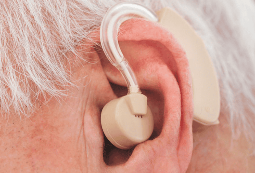
● Wear supportive metal devices such as: pacemakers, hearing aids, artificial joints, nails to fix broken bones...
In addition, patients will be asked to wear loose, comfortable clothes , no hospital lockers or changing clothes. They are also asked to leave the following items outside the studio:
● Cell phones;
● Dentures;
Eyewear;
Hearing aids;
Key;
Bras;
Wristwatches;
Wigs.
If the patient has claustrophobia, the patient may be familiarized with the machine first or given a sedative intravenously.

Khi chụp MRI, bệnh nhân được yêu cầu tháo máy trợ thính
3. MRI equipment
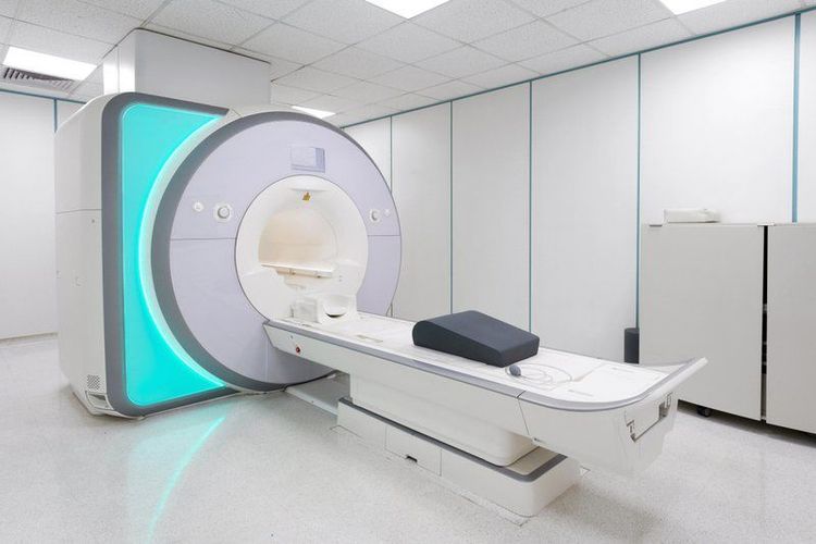
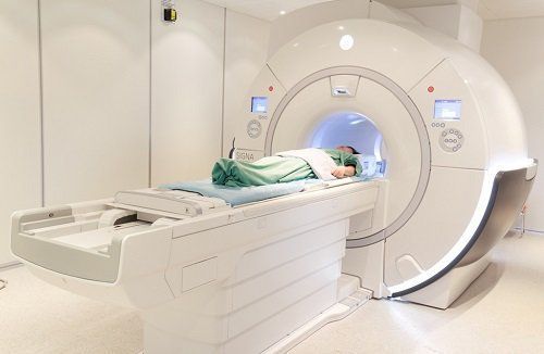
Magnetic resonance imaging machine is a large device with two ends surrounded by blocks of magnets. The patient will be asked to lie on a table that can be moved along the axis of the machine.
While taking the scan, the patient is not completely inside the system of the machine, but only the body part is assigned to be taken, the rest of the body is outside.
A machine called an open MRI is made for people with claustrophobia or excessive obesity. However, the image quality of open MRI machines is often not as good as that of closed MRI machines.

Máy chụp cộng hưởng từ tại Vinmec
4. What Happens During an MRI
Before the MRI, the patient will be injected with a contrast dye into a vein in the arm, usually gadolinium. This medicine gives the technician a better view of the structures inside the body. The patient will lie on an imaging table that moves along the body and is secured by a safety belt.
The MRI machine creates a strong magnetic field that affects the patient's body, the computer will pick up the signals from the protons in the body and present them as images through the technician's screen. Each primary photo is a thin slice of the body or area being taken.
During the scan, the patient can hear 2 loud sounds, that is, when the machine turns on and off the magnetic field, they will be provided with a silencer such as headphones or earplugs. Also, because MRI stimulates the nerves, mild seizures are common, but that's completely normal. The scan will take between 20 and 90 minutes depending on the size of the area being photographed.
In functional MRI, the patient may be asked to perform some simple tests such as touching the tips of the thumb to the tips of other fingers or answering questions asked by the doctor. This helps determine exactly which part of the brain controls these actions.

Bệnh nhân được tiêm thuốc cản quang trước khi chụp MRI
5. Who should not have an MRI?
The following cases are not indicated for MRI:
● Pregnant women in the first 3 months should not have MRI unless absolutely necessary. This is the time when the fetus's organs are developing, moreover, injecting contrast agents to the mother will also inadvertently pass them across the placenta into the baby's bloodstream.
● In case of allergy to contrast or severe kidney failure.
● People who wear metal support devices in the body.
6. Results to expect from an MRI scan
In case contrast injection is required, the technician will make sure to remove all the drug from the patient's body after conducting magnetic resonance imaging.
Allergic reactions occurring after injection of photosensitizers are very rare, however, you should immediately notify your doctor or technician if you experience any of the following symptoms: rash, hives, difficulty breathing ....
In case of claustrophobia and having to use sedatives, the patient will be asked to stay in a medical facility until he is completely awake or is taken home by his or her family.
Magnetic resonance imaging is becoming more and more popular because of the values it brings. There are many reasons why doctors assign their patients an MRI scan such as MRI of the brain, spinal cord, MRI of the heart, blood vessels, MRI of the abdomen... to help make an accurate diagnosis of the disease.

Chụp MRi giúp đưa ra chẩn đoán chính xác về tình trạng bệnh
Magnetic resonance imaging is not applied to pregnant women in the first 3 months, people with severe kidney failure as well as people wearing metal support devices because magnetic waves can adversely affect the above object. In addition, although allergies to contrast agents are rare, patients also need to be extremely vigilant when symptoms such as rash, hives, difficulty breathing... after an MRI scan appear.
Vinmec International General Hospital is currently one of the major hospitals with modern machinery and equipment for general medical examination and treatment procedures and accurate and modern MRI for brain and vascular diseases. brain blood in particular.
Especially, Vinmec International General Hospital is the first unit in Southeast Asia to put into use the new 3.0 Tesla Silent Resonance Imaging machine from the US manufacturer GE Healthcare.
The machine currently applies the safest and most accurate magnetic resonance imaging technology available today, without using X-rays, non-invasive. Silent technology is very beneficial for patients who are young children, the elderly, patients with weak health or have just had surgery.
Doctor Nguyen Dinh Hung has over 10 years of experience in the field of diagnostic imaging (Ultrasound, CT, MRI). Trained and practiced on hepatobiliary interventional radiology at Bach Mai Hospital (Intervention under ultrasound guidance, DSA, CT...) and deployed at the Diagnostic Imaging Department of Viet Tiep Hospital Hai Phong. Currently, he is a doctor at the Diagnostic Imaging Department of Vinmec Hai Phong International General Hospital.
To register for an examination at Vinmec International General Hospital, you can contact the nationwide Vinmec Health System Hotline, or register online HERE.
References: webmd.com, mayoclinic.org


