This is an automatically translated article.
The article was professionally consulted by Specialist Doctor I Nguyen Thanh Hai - Radiologist - Department of Diagnostic Imaging and Nuclear Medicine - Vinmec Times City International General Hospital.Cardiac magnetic resonance imaging (cardiac MRI) is a modern imaging technique that is being used commonly for patients with suspected heart disease, coronary artery disease, congenital heart disease, or congenital heart disease. heart failure, myocardium...
1. Overview
What is cardiac magnetic resonance imaging?Cardiac magnetic resonance imaging is a modern imaging technique in medicine that uses magnetic fields and radio waves to evaluate damage to heart structures such as pericardium, myocardium, chambers, and valves. , coronary artery system. Images from MRI have high contrast, good anatomical detail, allowing to detect lesions in terms of morphology and function of the heart (myocardial perfusion, tissue characteristics, coronary arteries).
Advantages of cardiac MRI:
Unlimited survey plane. High spatial resolution, high soft tissue contrast. Determine tissue characteristics, coronary angiography. Non-invasive diagnosis, high safety level. Does not cause side effects like CT or X-ray, still helps to detect abnormalities behind the bone layers. Imaging is faster and more accurate than X-rays in the diagnosis of cardiovascular disease. Disadvantages of cardiac MRI:
Movements in the heart, chest, diaphragm... can cause image noise. Structures smaller than 1mm in size will be difficult to examine (eg, coronary arteries). Examination time can be long, difficult to perform in patients with weak health who cannot lie still for a long time.

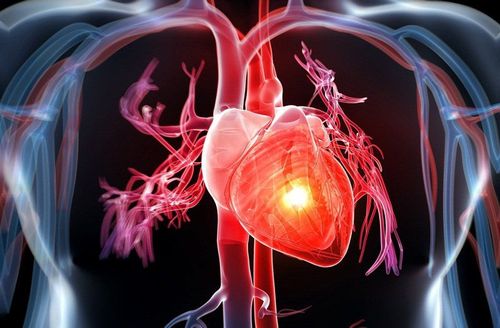
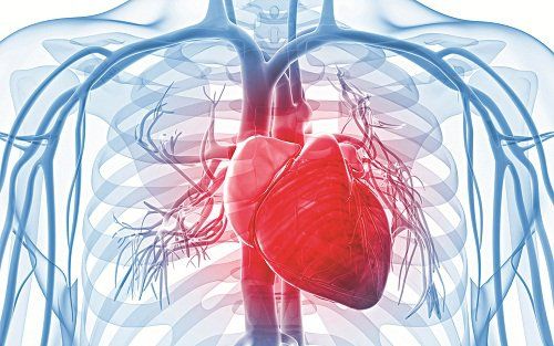
Chụp MRI tim giúp phát hiện các bất thường sau các lớp xương
2. Indications and contraindications
Indications for cardiac MRI for people with the following diseases:Cardiomyopathy: myocarditis, myocarditis, myocardial hypertrophy, myocardium with iron deposition, no raft... Congenital heart disease needs evaluation abnormality of flow, anatomy... Coronary artery disease: Evaluation of left ventricular function and mobility, assessment of myocardial survival in acute and chronic infarction. Heart valve disease: calcified heart valve, aortic regurgitation ... Pericardial disease: pericardial effusion, acute pericarditis Metabolic cardiomyopathy (Spectroscopy) Primary heart tumor, secondary heart tumor development: help provide images of the tumor, from which to diagnose and determine the extent of invasion.
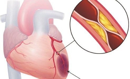
Bệnh nhân bệnh mạch vành cần chụp MRI tim
Patients wear electronic devices such as: cochlear implants, anti-vibration machines, pacemakers, Neurostimulators, automatic drug pumping devices under the skin.... Wearers of surgical forceps Intracranial, vascular, or orbital metal surgery <6 months (If worn for more than 6 months, consider further depending on the patient's health status). Severely ill people need resuscitation equipment on their side.
3. Cardiac magnetic resonance imaging procedure
3.1 Preparation for the procedure
To perform cardiac magnetic resonance imaging, it is necessary to prepare:Implementation team:
Electroradiologist. Optical technician. Means of use:
Resonant angiogram from 1.5 Tesla or more. Image storage system. Film printer. Movie. Supplies used:
18G venous puncture needle. Physiological saline/distilled water. 10ml syringe. Gauze, gloves, sterile bandages. Contrast drug accident first aid box.

Thuốc đối quang từ cần được chuẩn bị trước khi thực hiện thủ thuật
Magnetic Contrast Drugs. Sedative. Antiseptic for mucous membranes and skin. Patients need to prepare:
Be explained in advance about the procedure to coordinate well with the doctor. No fasting is required (if contrast is not required). Prepare a copy of the clinician's request for a scan, with a clear diagnosis or complete medical record (if necessary). Check for contraindications. Change into specialized clothes of the magnetic resonance imaging room according to the instructions, remove contraindicated items.
3.2 Implementation process
The whole procedure of cardiac MRI takes about 30 minutes in turn, following these steps:Patient position:
The patient is instructed to lie supine on the table. Select and position the receiver coil. Move the table into the machine compartment and locate the area to be photographed. Shooting technique
Positioning capture. Capture according to appropriate procedures depending on the pathology and clinical requirements. Taking pulse sequences helps to evaluate morphology and function on the level of 4 chambers, 2 chambers, short axis, right and left ventricular outflow tract... Injecting intravenous contrast to patients: Assessing muscle perfusion the heart is in rest ( Rest ) and in stress ( Stress ) if needed; evaluate the kinetics of contrast agents through the cardiac chambers on the pulse sequence (Dynamique). Note: Contrast injections during imaging may cause allergic reactions (urticaria, rash, nausea, anaphylaxis...) so emergency preparation is required in case of ear variable.
Contrast injection is taken in the late phase after 10 minutes, assessing myocardial survival. The technician processes the acquired images, prints the film, transfers the images and data to the doctor's workstation (The acquired image must clearly see the anatomical structure of the heart). Based on the above information, the doctor analyzes the lesion (if any) and makes a diagnosis.

Hình ảnh chụp MRI tim được thu lại và in phim
To protect cardiovascular health in general and detect early signs of cardiovascular disease, customers can sign up for Cardiovascular Screening Package - Basic Cardiovascular Examination of Vinmec International General Hospital. The examination package helps to detect cardiovascular problems at the earliest through tests and modern imaging methods. The package is for all ages, genders and is especially essential for people with risk factors for cardiovascular disease.
Doctor Hai has more than 20 years of experience in the field of diagnostic imaging, especially in the field of multi-segment computed tomography, magnetic resonance. Currently, the doctor is working at the Department of Diagnostic Imaging and Nuclear Medicine - Vinmec Times City International Hospital.
Customers can directly go to Vinmec Health system nationwide to visit or contact the hotline here for support.