This is an automatically translated article.
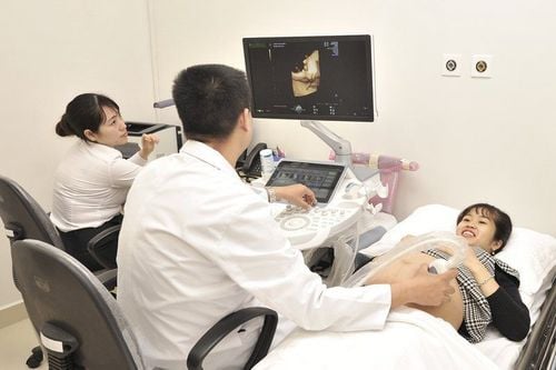
To monitor the development of the fetus, ultrasound is the most used method by pregnant women. Among ultrasound methods, super 3D technology offers good image quality, and the most accurate analysis results.
1. What are the benefits of 3D ultrasound?
3D ultrasound supports displaying volume of 3-dimensional image data showing the smallest details of the developing fetus in the womb.
In addition to checking the position of the fetus, the abnormal behavior of the fetus with 3D ultrasound helps the doctor detect fetal abnormalities.
2. What's the difference between 2D, Doppler, 3D and 4D ultrasounds?
2D (two-way) ultrasound uses a transducer placed on the abdomen or into the vagina to transmit sound waves through the body. The waves are emitted from the internal organs and fluids, and a computer converts these echoes into a two-dimensional (or cross-sectional) image of the fetus on a screen. With a Doppler fetal ultrasound, your doctor uses a handheld ultrasound device to amplify the sound of your baby's heartbeat with the help of a special cushion on your abdomen For a 3D ultrasound, multiple images dimensions are captured at various angles and then stitched together to form a three-dimensional rendering. 4D ultrasound is similar to 3D ultrasound, but shows moving images like a video. In the 4D ultrasound, you'll see your baby doing things in real time (like opening and closing eyes and sucking thumbs).

Siêu âm thai được chia thành nhiều loại khác nhau
3. When to perform 3D ultrasound?
Pregnant women with stable health, without unusual complications will have two 3D ultrasounds, while older mothers and those with complications will usually have more. Usually, there are many reasons to have a test done during pregnancy, depending on the trimester, including:
Confirm your expected date of transition. Monitor your baby's heart rate Make sure the pregnancy is not ectopic (ie in the fallopian tubes) and is in the uterus Confirm the number of babies in the uterus Ensures the baby is growing properly and at a suitable rate Check and measure your baby's major organs Measure your baby's size. Check amniotic fluid level Screen for birth defects Determine baby's sex Give parents a view of their baby and reassurance about their baby's normal development during pregnancy.
4. Image analysis during 3D fetal ultrasound
4.1 14-18 weeks 3D ultrasound will allow you to see the whole baby in one picture. Babies are very active during this stage of pregnancy. Therefore, if the baby is awake, you will be able to see all the movements of the fetus. You can watch your baby kick, wave, hold his feet, etc. During this time, you will be able to realize that your baby has two eyes, a nose and a mouth. However, the intricate detailed face composition will not be seen.

Từ 14 -18 tuần tuổi đã có thể nhìn rõ thai nhi cử động
4.2 19-26 weeks Baby's facial features will be more obvious. Therefore, doctors often recommend that you come for an ultrasound during this stage of pregnancy if you are pregnant with twins.
4.3 27-40 weeks In general, at this time, babies usually bow their heads. 27 to 40 weeks is the best time to get detailed close-up photos of your baby's face in 3D ultrasound.
In all stages of pregnancy, the most important factor affecting the quality of 3D ultrasound images is the amount of water that the mother takes into her body in the period near the visit date. The right amount of water will ensure that there is enough fluid in the amniotic sac to see the baby in detail.
5. Some notes that mothers need to know when having a 3D pregnancy ultrasound
If you are under 12 weeks pregnant, you should drink water, about 2-3 glasses of water about an hour before the ultrasound and must hold your urine. This extra water will stretch the bladder and make it easier to have a 3D pregnancy ultrasound.
For fetuses over 12 weeks, pregnant women need to urinate completely to perform ultrasound. In addition, some other cases will follow the doctor's instructions.
Although ultrasound does not affect the fetus, pregnant women are also very careful not to do too many ultrasounds during pregnancy because this does not bring many benefits, but also wastes time and money for pregnant women. family.
3D fetal ultrasound will help doctors see the development of the fetus as well as detect any abnormal signs, if any, to promptly intervene with appropriate methods.
However, it is not except for cases where a doctor can detect birth defects until birth.
For all information about maternity and childbirth services at Vinmec Hospital, please contact the hospitals and clinics of Vinmec Health system nationwide.
Please dial HOTLINE for more information or register for an appointment HERE. Download MyVinmec app to make appointments faster and to manage your bookings easily.