This is an automatically translated article.
3D echocardiography is very meaningful in the assessment and intervention of heart valve diseases. 3D ultrasound is more accurate than 2D ultrasound, but requires a place equipped with modern and high-level machines.
1. Learn about 3D echocardiography in the assessment and intervention of valvular diseases
Echocardiography uses ultrasound waves to reconstruct the heart's chambers, the walls of the heart valves, parts of the heart valves, and the great blood vessels. Techniques on 2D ultrasound to provide the most valuable classical parameters for the diagnosis and treatment of cardiovascular diseases without invasiveness, only through the chest wall or minimally invasive. as minimal as transesophageal ultrasound.
Advances in computer and transducer technology over the past two decades have allowed the development of three-dimensional (3D) echocardiography (3DE), providing significant additional clinical information to two-way echocardiography. traditional (2D) dimension.
Through many recent studies, 3D echocardiography has been shown to be useful for clarifying complex cardiac anatomy and hemodynamics of the intracardiac circulatory system. The recently introduced real-time 3D echocardiogram can overcome technical and quality issues and is aimed at clinicians learning the widespread use of 3D echocardiography in clinical settings. routine clinical basis. 3D echocardiography can provide visual recognition of cardiac structures from any spatial orientation and can provide complete information about the absolute volume and function of the cardiac chambers and methodological trends. This will be one of the ultimate goals of cardiac imaging.
2. Indications of 3D echocardiography in the assessment and intervention of valvular diseases
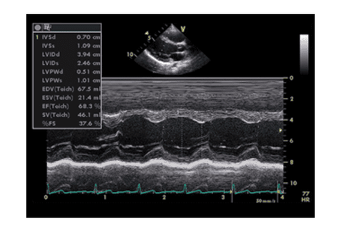
3D echocardiography increases the ability to survey the heart wall: thickness / thinness, mobility of the heart wall, thereby providing information about the profile picture in ischemic heart disease. Echocardiography can be used to evaluate left ventricular (LV) systolic function as well as left ventricular diastolic function. From there, it can help diagnose, evaluate function or evaluate the effectiveness of treatment in many diseases such as left ventricular hypertrophy, hypertrophic or restrictive cardiomyopathy, severe heart failure, pericarditis. Heart spasm... Similarly, surveying right ventricular volume (RV) by 2D ultrasound requires geometric modeling, which is particularly difficult due to the crescent shape of the right ventricle. And clearly 3D ultrasound is superior to 2D, making images easier to survey, more accurate or technically simpler.

Siêu âm tim 3D được chỉ định đánh giá và can thiệp các bệnh van tim
3D echocardiography also helps evaluate the size, function, or pressure of the great blood vessels: aorta, pulmonary artery, vena cava...
3. Advantages of 3D echocardiography in assessment and intervention of valvular diseases
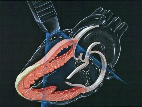
The information needed for this work was previously performed primarily with transthoracic or transesophageal 2D ultrasound, but now with 3D echocardiography offers significant advantages when evaluating mitral valves and heart valve repair plan. Recently, the properties and advantages of 3D ultrasound have greatly helped to assist in percutaneous mitral valve repair with the MitraClip device. It has been shown to overcome significant limitations with 2D ultrasound, which can only provide a limited assessment of anatomical and morphological changes during and after percutaneous mitral valve repair. In general, there are two main advantages of 3D imaging over conventional 2D echocardiography as follows:
Quantification of absolute cardiac chamber volume, including left ventricular (LV), right ventricular (RV) volume and the left atrium (LA) and their function. 3D structural images and moving images of the heart, especially the structure of the heart valves. To protect cardiovascular health in general and detect early signs of cardiovascular disease, customers can sign up for the Cardiovascular Screening Package of Vinmec International General Hospital. The examination package helps to detect cardiovascular problems at the earliest through tests and modern imaging methods. The package is for all ages, genders and is especially essential for people with risk factors for cardiovascular disease.
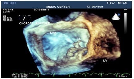
Siêu âm tim 3 chiều phát hiện đứt dây chằng van 2 lá
Why should you choose Cardiovascular Screening Package at Vinmec International General Hospital?
Simple and quick procedure. Enthusiastic advice and support, reasonable and convenient examination process. Comprehensive facilities, including a system of clinics and consultations, blood collection rooms, dining rooms, waiting areas for customers... Professional, caring way of working.
Please dial HOTLINE for more information or register for an appointment HERE. Download MyVinmec app to make appointments faster and to manage your bookings easily.