This is an automatically translated article.
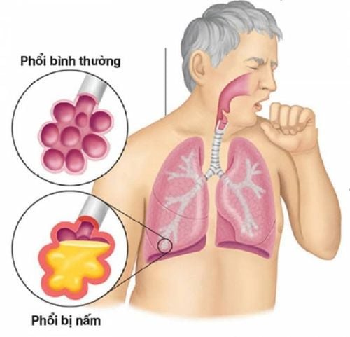
Lung fungal infection is a common lung infection in the elderly or immunocompromised, chronic disease. There are 3 common types of lung fungi: Aspergillus, Candida and Cryptococcus. The following article shares about the symptoms, diagnosis and treatment methods of the fungus in the lungs that often causes disease.
Although accounting for a very low rate in lung diseases, lung fungus if not detected and treated promptly can lead to high mortality.
1. Lung fungus Aspergillus
Aspergillus causes lung fungal disease in 3 types:
Lung mycosis
1.1 Symptoms
Lung fungal tumor: There may be one or more symptoms such as high fever (from 39 - 40 degrees Celsius), coughing up blood (which can be severe and very recurrent), chest pain, weight loss, fatigue. Mycosis fungoides have a higher risk of occurrence in patients with pulmonary tuberculosis, HIV, cancer chemotherapy patients, patients using corticosteroids and or long-term immunosuppressive drugs. Allergic bronchopneumonia: This is a condition in which the bronchopneumonia increases susceptibility to the fungus, with symptoms of persistent, long-lasting asthma, bronchial asthma that develops in episodes in patients with sinusitis, rhinitis. Allergies, eczema, food allergies, ... Infiltrative lung fungus: Usually occurs in immunocompromised patients or reduced white blood cell count, with one or more symptoms such as fever, persistent dry cough , mild to severe hemoptysis, chest pain, loss of appetite, weight loss, fatigue.
1.2 Diagnosis
Lung fungal tumor: Chest X-ray showed a rattle-like lesion with a solid mushroom mass and signs of gas sickle on the fungal mass. CT-scan shows that in addition to the rattle-shaped lesion, it may also be accompanied by other lesions or not, such as pleural thickening, calcification in the lung fungal mass, cocoon-like degeneration, and retractive pulmonary fibrosis. Performing microbiological tests such as direct examination or sputum culture, ... can identify Aspergillus fungus in the lungs. The diagnosis of lung mycoma must be differentiated from lung abscess and abscessed lung cancer. Allergic bronchopneumonia: X-ray shows large bronchi with dilated bronchi. Blood tests showed increased eosinophils and IgE levels in the blood. Performing sputum culture can find Aspergillus fungi and eosinophils. The diagnosis of allergic bronchopulmonary Aspergillus should be differentiated from obstructive fungal bronchitis, bronchial granulomatosis, eosinophilic pneumonia, and parasitic lung disease. Infiltrative pulmonary fungal: X-ray and CT-scan of the lung show pulmonary lesions as clumps or opacities, around the nodule with a shadow (bleeding), which may or may not be associated with necrotizing pneumonia. , abscess drive. Other lesions may be accompanied by effusion, pneumothorax. Bronchoscopy showed lesions in the tracheobronchial lumen with ulcerative white pseudomembranous lesions (occasionally). If sputum culture is found, Aspergillus fungus is found.
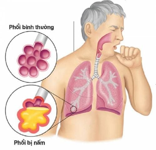
Phổi nhiễm nấm có thể được chẩn đoán bằng phương pháp chụp X-quang phổi
1.3 Treatment
Lung fungal tumor: Treatment is by surgical method of removing a lobe of the lung or removing one lung. For lung fungal tumors, the use of antifungal drugs has little effect. In case the patient cannot be operated on and has severe hemoptysis, bronchial artery occlusion is indicated. Allergic bronchopneumonia: The basic treatment with allergic bronchopneumonia is using oral corticosteroids to reduce allergies and hypersensitivity of the lungs to the fungus. Infiltrating lung fungus: Depending on the condition of the lung fungus infiltrating the patient and the conditions at the medical facility, choose to use one of two antifungal drugs, Amphotericin B-Deoxycholate or Itraconazole.
2. Candida
2.1 Symptoms
Candida pneumonia is common in patients with weakened immune systems, long-term decreased white blood cell count, patients being treated with intravenous catheters or catheters. Symptoms of the disease may be as follows:
Moderate to severe fever, sudden and prolonged fever Cough with phlegm, dry cough, possibly hoarseness White tongue causing difficulty swallowing, ulcers with false white on the mucous membranes of the mouth and throat Chest pain, sternum, shortness of breath, wheezing
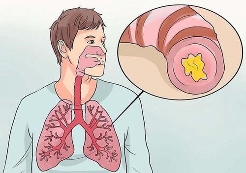
Ho ra đờm là một dấu hiệu nấm phổi
2.2 Diagnosis
Chest X-ray shows a fungal infection of the lung with clumps or opacities. Round nodules, maybe one or more nodes located in different places in each cluster. Bronchoscopy showed that the surface of the bronchial lumen was damaged by a non-adhesive, grayish-colored layer and in some cases accompanied by necrosis on the bronchial mucosa. Endoscopy of the esophagus and stomach can find Candida fungi causing some lesions. Blood cultures found Candida in the blood.
2.3 Treatment
Treatment of Candida lung fungus is mainly using the antifungal drug Amphotericin B for 4-6 weeks in combination with flucytosin cytosine. During treatment, pay attention to monitor the patient's electrolytes to promptly supplement potassium. If the esophagus is also infected with Candida, it is necessary to combine treatment with Ketoconazole and Fluconazole or Itraconazole for 2 weeks.
3. Cryptococcus
3.1 Symptoms
Cryptococcus lung fungus causes the following symptoms:
Fever, dizziness, headache Consciousness disorder, convulsions, possibly even coma, motor paralysis, nerves Persistent cough, shortness of breath, chest pain Appearance of blisters and ulcers on the skin
3.2 Diagnosis
Chest X-ray showed a uniform infiltrative lung fungal lesion, with enlarged lymph nodes in the hilum, collapsed lobes or lobes, and bronchiectasis. The lung parenchyma is injured in the form of a reticular nodule, lung abscess or pleurisy. Blood tests showed increased IgE levels. Precipitin quantitative assay. Test for Cryptococcus by staining with Chinese ink or culture.
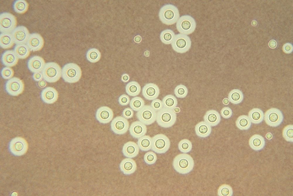
Hình ảnh nấm phổi Cryptococcus
3.3 Treatment
Treatment of Cryptococcus depends on the patient's condition:
Severe Cryptococcus pneumonia (not associated with HIV): Initial treatment with Amphotericin B for 4 weeks in combination with Flucytosin, then consolidation with Fluconazole for 8 weeks the week. Mild Cryptococcus: Treat with fluconazole for 6-12 months.
4. Prevention of lung fungal diseases
Lung fungus, if not detected and treated promptly, can create conditions for fungal bacteria to move to other organs causing disease such as skin, muscle, meninges, blood vessels... leading to infection, body weaken or leave serious complications.
Therefore, it is very important to prevent lung fungal infections with measures such as:
Eat a nutritious diet, especially vegetables and fruits to increase resistance, improve the immune system, combine exercise sports. Keep house hygiene, clothes dry, clean, sunny to avoid mold. To prevent 3 common and disease-causing fungi in the lungs, Aspergillus, Candida and Cryptococcus, we need to pay attention to eating and exercising to maintain the health of the immune system, and at the same time, avoid places, activities, and furniture. For personal use with mold. In particular, periodic health check-ups play an extremely important role, through which the doctor will check the overall health to detect the causes and signs in order to have timely intervention.
General health check-up package - Vinmec International General Hospital is deployed for all customers and ages. All medical examination procedures are performed by a team of doctors with many years of experience. Therefore, customers can be assured of medical quality at Vinmec.
Please dial HOTLINE for more information or register for an appointment HERE. Download MyVinmec app to make appointments faster and to manage your bookings easily.


