This is an automatically translated article.
With the fact that cardiovascular disease is increasingly common in modern times and the risk of death is high, nuclear medicine is holding a certain role in the diagnosis of cardiovascular diseases thanks to advantages such as: no invasion, providing a wealth of morphological and functional information as well as anomaly detection with high sensitivity. Currently, there are three most common types of cardiovascular scans: myocardial perfusion scan, ventricular scintigraphy, and myocardial scar marking scan.
1. What is myocardial perfusion scan?
Myocardial perfusion scintigraphy is a method of using intravenous radiopharmaceuticals to evaluate the myocardial absorption of these substances, and is proportional to the degree of perfusion. Therefore, the areas of myocardium that are less radioactively attached are also the areas of partial or complete hypoperfusion
In addition, myocardial perfusion scintigraphy also helps to compare the radiopharmaceutical absorption status of the muscle. the heart is in the exercise phase versus the stationary phase. However, this comparison is also influenced by factors such as the degree of coronary stenosis, the hemodynamic effects of the stenotic coronary artery, the capacity of the coronary flow reserve (exercise-static) and The number of coronary arteries is narrowed
However, in some cases, the decrease in myocardial activity can also be a false positive due to soft tissue occlusion, mammary glands in women reduce the activity image or other abnormalities. Diaphragm and abdominal viscera reduce the activity of the lower wall myocardium on scintigraphy.

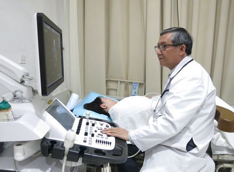
Xạ hình tưới máu cơ tim đánh giá khả năng hấp thụ dược chất phóng xạ của cơ tim
Myocardial perfusion scintigraphy is also performed along with exercise testing to:
Diagnose the cause of chest pain in patients with unknown diagnosis Determine functional impact of the stenotic branch of the coronary artery Identify functional role of the collateral coronary branches Assess the effectiveness of coronary reperfusion after coronary artery bypass surgery, percutaneous coronary intervention or fibrinolysis Prognosis of survival after myocardial infarction heart
2. What is a ventricular scintigraphy?
Ventricular scintigraphy is a method of imaging to evaluate the size, shape and movement of each region of the ventricles at various stages of the cardiac cycle. This method is very valuable in evaluating the ejection fraction at rest and during exercise in patients with coronary artery disease, valvular heart disease, or congenital heart disease. In addition, scintigraphy can also be used for long-term monitoring of ventricular function in patients receiving cardiotoxic chemotherapy for cancer. However, ventricular scintigraphy has now been largely superseded by echocardiography, a less expensive technique that avoids radiation exposure.

Xạ hình tâm thất đồ giúp đánh giá phân số tống máu khi nghỉ và khi gắng sức ở bệnh nhân
Based on first absorption phase analysis Based on multi-minute synchronized electrocardiogram (MUGA) Indications for Ventrographic scintigraphy includes:
Determination of total ventricular function (T) and prognosis of ischemic heart disease Determination of regional movement disorders in patients with ischemic heart Determination of ventricular function ( T) in patients with ischemic cardiomyopathy Monitor patients after treatment with Anthracyclines Determine cardiac function before and after heart transplantation Determine ventricular size and function (T) in patients with valvular disease Determine Determine the ability to preserve left ventricular function during exercise in patients with coronary artery disease and patients with aortic regurgitation Determine right ventricular function in pathologies such as acute inferior myocardial infarction, bronchiectasis , congenital heart disease, left ventricular failure, arrhythmia due to right ventricular dysplasia

Kỹ thuật xạ hình tâm thất đồ hiện nay hầu như đã bị thay thế bởi siêu âm tim
3. How does a scan mark myocardium scar?
After myocardial infarction, myocardial perfusion scintigraphy has prognostic value to help assess the area of ischemia, the degree of scarring of the old infarct and the scarred areas of the infarct or other areas, but also the possibility of Reperfusion
Specific infarct scintigraphy also helps to detect infarct scar areas at about 12-24 hours up to 1 week after acute myocardial infarction. If there is progressive necrosis of the myocardium after myocardial infarction or progressive ventricular aneurysm, the scan results can also detect
Cardiovascular Center - Vinmec International General Hospital owns a team A team of experts including professors, doctors, specialists 2, and masters are experienced, have great reputation in the field of medical treatment, surgery, interventional cardiac catheterization and application of advanced techniques. in the diagnosis and treatment of cardiovascular diseases. In particular, the Center has modern equipment, on par with the most prestigious hospitals in the world.
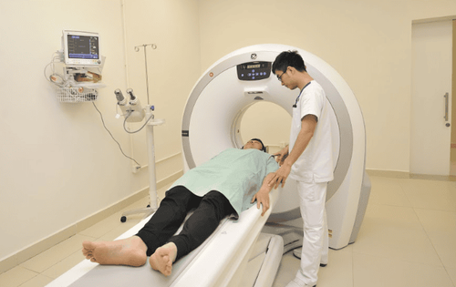
Vinmec Hospital uses the SPECT/CT Discovery NM/CT 670 Pro system with the most modern 16-sequence CT of the world's leading medical equipment company GE Heathcare (USA), for high quality images, helping to diagnose early disease to investigate. Vinmec is one of the few hospitals implementing routine myocardial perfusion scintigraphy. Experienced, well-trained Vinmec medical staff at home and abroad will provide high quality services and maximum support for customers.
If you have a need for consultation and examination at Vinmec Hospitals under the national health system, please book an appointment on the website for service.
Please dial HOTLINE for more information or register for an appointment HERE. Download MyVinmec app to make appointments faster and to manage your bookings easily.


