This is an automatically translated article.
This article is professionally consulted by - Master, Resident, Specialist I Trinh Le Hong Minh - Department of Diagnostic Imaging - Vinmec Central Park International General Hospital.X-rays are known to be one of the important discoveries in the history of modern medicine. The invention of X-rays has brought great applications to help detect and support diagnosis and treatment of diseases with high efficiency and accuracy.
1. What are X-Rays?
The nature of X-rays is an electromagnetic wave, the radiation emits a beam of electrons hitting a solid object, most X-rays have a wavelength range between 0.01 and 10 nanometers corresponding to the frequency range from 30 Petahertz to 30 Petahertz. 30 Exahertz (3×10¹⁶ Hz to 3×10¹⁹ Hz) and has energies from 120 eV to 120 keV. The wavelength of X-rays is longer than gamma rays but shorter than ultraviolet rays.During the 19th century, X-rays were considered as one of the outstanding inventions. Not only opening a new chapter for physics, but X-rays are also widely used in the medical field, helping doctors to see the internal parts of the patient's body without surgery.
In 1985, physicist Wilhelm Conrad Rontgen (1845-1923) born in Lennep, Germany, became the first person to observe X-rays, this is also why X-rays are also called Rontgen rays. This is considered an important scientific invention that is applied in many different fields, mainly medicine.
X-rays are a form of high-energy radiation that cannot be seen with the naked eye. The application technique of x-rays is widely used in many fields, especially in medicine, because this type of ray can penetrate many objects, especially living ones. However, X-rays also bring many dangers to the human body.

Tia X là một dạng bức xạ năng lượng cao không thể nhìn thấy được bằng mắt thường
2. X-ray penetration
X-rays, an electromagnetic radiation, are emitted by electrons located outside the nucleus of an atom (unlike other high-energy alpha and gamma rays, which are emitted from the nucleus of an atom). X-rays are similar to visible light but have much shorter wavelengths (from about 10 to 0.02 nanometers) and much higher energies (from ~0.12 to ~120 keV). Thus, X-rays can penetrate biological tissues and many other materials that visible light cannot. This high penetrating power and different X-ray attenuation coefficients of different body tissues make X-rays a useful signal for biomedical imaging.In general, X-rays are classified as either soft or hard according to their energy range. Soft X-rays range from ~0.12 to ~12 keV and hard X-rays range from ~12 to ~120 keV. Understandably, hard X-rays are generally used for solid or large objects, and soft X-rays are used for small objects or for some special requirements for low-energy imaging.
X-ray imaging depends not only on the X-ray energy, but also on the density of the materials to be imaged; The higher the density of the material, the less absorption and less penetration. It is because of these differential absorptions (i.e. X-ray attenuation coefficients) that different densities of bone, muscle, fat and other soft tissues can be distinguished. This is the physical basis of biomedical radiographic imaging.
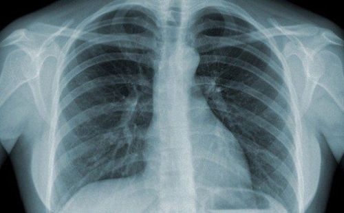
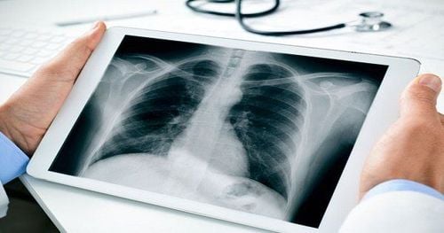
Because of the differences between the X-ray attenuation coefficients of the different tissues that produce the image contrast, X-ray-based technologies are generally good for imaging bones and lungs. Because bone contains relatively heavy atoms with many electrons that act as X-ray absorbers, its appearance is significantly different from that of surrounding soft tissues, which are mainly composed of water, proteins, and molecules. Others have lighter atoms.
In the lungs, the air does not absorb X-rays, so it acts as a contrast agent, the structure of the lung tissue can be clearly seen due to the difference in contrast between the air and the lung tissue. . However, for soft tissues and organs, radiographic images produce very poor contrast. Because of this lack of contrast, X-ray-based techniques may not be ideal for soft tissue and organ imaging without the use of contrast media.
The shorter the X-ray wavelength, the greater the penetration. It easily passes through objects that are not transparent to ordinary light such as wood, paper, fabric, soft tissues such as meat, skin. For hard tissues such as bone and metal, it is more difficult to pass. The larger the atomic mass of a metal, the harder it is for X-rays to penetrate. For example, an X-ray beam can pass through a sheet of aluminum several centimeters thick, but be blocked by a sheet of lead several millimeters thick. Therefore, lead is often used as a protective shield in the X-ray room.
Tia X có thể đi qua các vật không trong suốt với ánh sáng thông thường
First, you consider where the X-ray tube is going, and what rooms are on the other side of those walls (including floor/ceiling). Then you factor in what those rooms are used for and how often they are used. Finally, you consider how powerful the X-ray equipment is and how often it is used. You combine all of that to figure out how much shielding you need to keep exposure within safe limits.
3. Applications of X-Rays
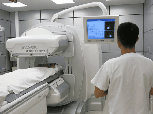
Unlike infrared rays, which are widely used in everyday life, X-rays are used a lot in medicine to take X-rays, treat cancer and many other purposes including:X-ray images This method helps doctors identify Determine if you have any joint problems. If the bone is damaged, such as broken or cracked, an X-ray will help detect the exact location of the affected bone. Computed tomography or CT scan is also an important application of X-rays.
Besides, X-rays also play an important role in checking transport security for goods, luggage and passengers. Real-time content of packages and guests' faces are displayed on electronic image detectors.
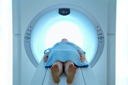
Radiation therapy for cancer By using high-energy radiation, radiation therapy destroys cancer cells by damaging their DNA. This treatment can also damage normal cells.
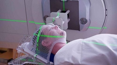
Tia X được ứng dụng trong xạ trị ung thư
This can damage or destroy cells if appropriate. It can also be used against it in cases where these cell damage could cause cancer. It can destroy those abnormal cells by directing X-rays at malignant tumors.
In modern medicine, X-rays play an extremely important role, but our health can be directly affected if we are exposed to a lot of X-rays.
X-rays today are widely used. medical field to treat cancer. The National Cancer Institute recommends that treatment be carefully planned to minimize side effects because X-rays also damage normal cells.
X-rays can be used to identify holes and cracks inside metal castings, or to study the structural composition of solids in the laboratory due to their ability to penetrate certain materials certain.
In the field of astronomy, X-rays are produced by certain celestial bodies in the universe, such as binary star systems or black holes at the centers of spiral galaxies that are swallowing stars and surrounding gas clouds . Therefore, scientists can use X-ray telescopes to study them.
Before taking a job at Vinmec Central Park International General Hospital, the position of Doctor of Radiology since February 2018, Doctor Trinh Le Hong Minh used to work as a resident in the Radiology Department at Vinmec Central Park. hospitals: Cho Ray, University of Medicine and Pharmacy, Oncology, People's Gia Dinh, Trung Vuong... from 2012-2015. Working officially at Cho Ray Hospital from 2015-2016, City International Hospital since 2016.
Please dial HOTLINE for more information or register for an appointment HERE. Download MyVinmec app to make appointments faster and to manage your bookings easily.
Reference sources: sciencedirect.com, reddit.com
SEE MORE
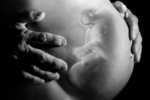
Does X-ray have any effect on health? 22 frequently asked questions about X-rays Effects of X-rays on the fetus