This is an automatically translated article.
Surgery for nerve tumors in general and surgery for meningiomas in particular by the method of opening the skull cap is a delicate operation that requires a high level of expertise, which should be carried out according to the correct procedure and according to Close postoperative follow-up.
1. Introduction to meningiomas of the occipital foramen
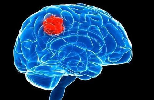
Meningioma is a fairly common intracranial tumor, originating from the arachnoid membrane (the thin membrane covering the brain and spinal cord parenchyma, different from the dura). Although most meningiomas are benign and slowly progressing, they can grow to very large sizes until detected. Several types of meningiomas are found along the dura mater in the venous sinuses of the brain and on the floor of the skull, where there are many capsular cells in the arachnoid layer.
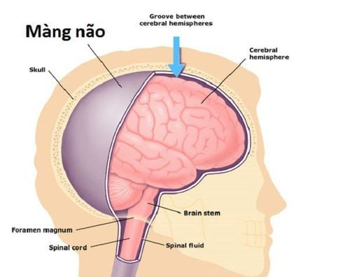
Occipital meningiomas occur near the opening of the base of the skull from which the lower part of the brain stem descends. Meningiomas of the occipital foramen are classified as quite rare, to access the tumor may require surgery to open the middle cranial fossa or posterior cranial fossa.
Manifestations of meningiomas progressing very slowly. Patients often present with headaches and signs of damage to various cranial nerves. In the early stages it is quite difficult to develop clinically.

Bệnh nhân bị u màng não lỗ chẩm có ít triệu chứng
2. Why surgery for meningiomas of the occipital foramen?
Surgery is considered an effective treatment for symptomatic meningiomas. Occipital foraminal meningioma surgery will be indicated in the following cases:
Occipital meningioma is dominant posteriorly. Tumor volume extending into the posterior fossa.
3. Prepare for surgery
Before the surgery, the patient will have a clinical examination, specialized evaluation of ophthalmology, endocrinology, liver and kidney function tests, blood coagulation, otolaryngology, brain magnetic resonance imaging, and imaging. CT assessment of skull base bone structure, meningioma of occipital foramen.
Then the patient was washed... When brought into the operating room, the patient was instructed to keep his head 30 degrees, straight, not tilted or bent.
The composition of the operating team includes 7-8 people including anesthesiologists, neurosurgeons, surgical assistant doctors, anesthesiologists, nurses serving surgical instruments, Nursing.
Surgical instruments include skull cap opener, microsurgery glasses capable of recording during surgery, Sonopet ultrasonic scalpel, microsurgical scissors, hemostatic device, hemostatic glue, stimulator monitor neural electrical activity...

Bác sĩ chuẩn bị dụng cụ trước khi tiến hành phẫu thuật
4. Surgical procedure for meningioma of the occipital foramen
Step 1: When preparing for surgery, the patient is placed in the prone position.
Step 2: Anesthetize with a mixture of Adrenalin 1/1000 solution and Lidocaine; followed by endotracheal anesthesia.
Step 3: The surgeon makes a skin incision right in the middle of the patient's posterior fossa to open the occipital bone and expose the tumor.
Step 4: Place microsurgery glasses to record during surgery, open the dura mater to aspirate cerebrospinal fluid.
Step 5: Remove the tumor piece by piece with a suction machine or ultrasound knife.
Step 6: Close the dura, close the incision in all 3 layers: fascia, skin, and subcutaneous.
5. Monitoring and handling postoperative complications
Postoperative monitoring
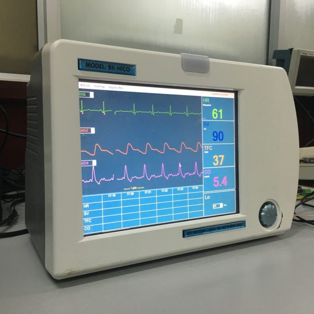
Monitor the patient's pulse, respiratory, temperature, pupil, blood pressure, anesthetic release time, consciousness, liver and kidney function... of the patient to assess vital signs survival, surgical results. Monitor complications and complications (if any).
Theo dõi huyết áp bệnh nhân đề phòng biến chứng sau phẫu thuật
Handling complications
After surgery for meningiomas, some risks may still occur, leading to complications. Some common complications after surgery may include:
Bleeding (intraoperative): The surgical process may inadvertently affect the veins leading to intraoperative bleeding. In this case, Surgicel can be used to stop bleeding or consider intravenous cauterization. Bleeding (after surgery): Often causes patients to be mentally retarded, difficult to get out of anesthesia or not awake for a long time, with focal neurological signs. Film should be taken for evaluation. To handle, it is possible to treat conservatively or drain the ventricles if there are signs of ventricular dilatation. Hematoma on the tentacle: Usually occurs due to the decrease in intraoperative pressure, patients with hematoma will show signs of impaired consciousness. In this case, it is necessary to consider the size of the hematoma to choose the surgical direction or medical treatment. Cardiovascular complications (intraoperative): During surgery for meningiomas, patients may present with tachycardia or hypertension due to brainstem stimulation. To improve the situation, the doctor can take out the decompression pad and combine it with pain relievers from the anesthesiologist. Ventricular dilation: A condition in which cerebrospinal fluid is blocked and accumulates too much (possibly due to bleeding or hemorrhage). The only solution is ventricular drainage. Meningitis: The patient will have complications of meningitis through fever, cerebrospinal fluid, bacteria, and increased white blood cells. For treatment, it is necessary to use antibiotics according to the antibiogram and actively treat. Vinmec International General Hospital with a system of modern facilities, medical equipment and a team of experts and doctors with many years of experience in neurological examination and treatment, patients can completely rest in peace. examination and treatment center at the Hospital.
To register for examination and treatment at Vinmec International General Hospital, you can contact Vinmec Health System nationwide, or register online HERE.


