This is an automatically translated article.
This article was professionally consulted by a Doctor of Radiology, Department of Radiology - Vinmec Central Park International General Hospital.Color Doppler echocardiography is a very valuable modern imaging tool in the diagnosis of cardiovascular diseases, especially heart valve diseases. Examination of the mitral valve is significant in assessing the extent and selection of treatment methods.
1. What is color Doppler ultrasound?
Color Doppler ultrasound is a modern form of ultrasound with the highest accuracy and easy-to-observe color ultrasound images. This is a diagnostic imaging method for the purpose of assessing the state of activity, structure of the heart or the state of blood movement in the circulatory system, hemodynamic status. From there, detect signs or abnormal diseases of the cardiovascular system. Therefore, this is a method that is prescribed and used by many doctors in the process of diagnosing and evaluating cardiovascular diseases. In addition, color Doppler ultrasound is also indicated in a number of other diseases.
2. Learn about mitral stenosis
Mitral stenosis is a very common disease in our country, often due to sequelae of rheumatic heart disease. After several episodes of recurrent rheumatic fever, mitral stenosis begins to develop, which continues to progress for many years until symptoms appear.
Hẹp van hai lá là bệnh rất phổ biến ở nước ta
The main lesions in mitral stenosis are fibrous infiltration, valve leaflet thickening, margin adhesions, adhesions and contracture of ligaments and muscle columns that contribute to mitral stenosis, calcification appears on the leaflets, ligaments, annulus, limit the normal function of the heart valve for a long time leading to heart failure.
In order to accurately diagnose mitral stenosis and the degree of stenosis for timely treatment, color Doppler ultrasound should be performed.
3. Evaluation of mitral stenosis on color Doppler echocardiography
Objectives of color Doppler echocardiography in mitral stenosis:
Determine and diagnose the degree of mitral stenosis, Features of the leaflets, annulus, margin and subvalve apparatus Area of valve opening Cardiac chamber size and circulating blood flow or left atrial blood clot Pulmonary artery pressure Left ventricular size and function Injury to other heart valves, other heart disease in combination Specific:
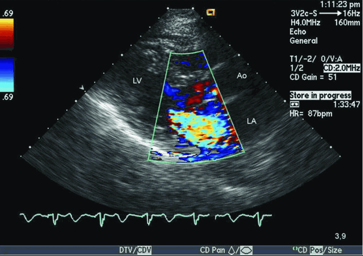
Color Doppler ultrasound helps to evaluate Peak mitral flow velocity > 1 m/s suggests mitral stenosis, but this finding is nonspecific and can be caused by tachycardia, increased contractility, mitral regurgitation, and ventricular septal defect. The transmural gradient (proportional contouring of the mitral flow) allows an estimate of the severity of the stenosis. Mild mitral stenosis: mean transvalvular pressure difference < 5 mmHg, moderate mitral stenosis: mean valvular pressure difference 5-12 mmHg constricted mitral stenosis: mean transvalvular differential pressure > 12 mmHg.

Hình ảnh siêu âm tim Doppler màu để đánh giá hẹp van hai lá
Color Doppler ultrasound is used to estimate pulmonary artery pressure by measuring the spectrum of associated tricuspid regurgitation or associated pulmonary regurgitation seen in mitral stenosis. Allows assessment of the associated damage such as mitral regurgitation, aortic regurgitation and extent, which is very important to help decide on the appropriate mitral valve intervention method. Doppler echocardiography is used to assess the area of the mitral orifice quite accurately, thereby identifying and evaluating the degree of stenosis. Mitral stenosis is very tight when the orifice area is < 1.0 cm2. Mitral stenosis is tight when the orifice area is from 1 to < 1.5 cm2. Mitral stenosis is moderate when the area of the orifice is 1.5-2.0 cm2. PHT (half-pressure-relief time) method: The depressurization half-life (time for the pressure to decrease to half from its initial value), is the time for the velocity to decrease to 70% of the peak velocity. Mitral stenosis prolongs the decompression time of flow through the mitral valve. The narrower it is, the longer this period is.
4. What are the advantages of color Doppler ultrasound?
Color Doppler ultrasound is a modern type of ultrasound, widely used, with many outstanding advantages such as:
Ultrasound is easily performed many times with a fairly moderate cost. Ultrasound is considered safe, does not affect the whole body and causes no pain or discomfort.

Siêu âm Doppler màu là loại siêu âm hiện đại, được ứng dụng phổ biến
High accuracy makes it easier for doctors to diagnose and better detect diseases Ultrasound has the ability to detect situations that can occur in hemodynamics, hemodynamics, Mode B anatomy that other forms of ultrasound do not can be done. However, this is a high technique that requires the experience and expertise of the doctor reading the results. Therefore, the effect can only be achieved in highly qualified medical facilities. Patients can be completely assured when choosing to evaluate mitral stenosis on color Doppler echocardiography at Vinmec International General Hospital, not only having a system of modern facilities and medical equipment, Vinmec also owns a team of experts and doctors with many years of experience in the examination and treatment of cardiovascular diseases. In particular, customers can register for Cardiovascular Screening Package - Basic Cardiovascular Examination of Vinmec International General Hospital. The examination package helps to detect cardiovascular problems at the earliest through tests and modern imaging methods. The package is for all ages, genders and is especially essential for people with risk factors for cardiovascular disease.
Please dial HOTLINE for more information or register for an appointment HERE. Download MyVinmec app to make appointments faster and to manage your bookings easily.


