This is an automatically translated article.
Fetal doppler ultrasound is a common imaging technique in obstetrics to examine the fetal heart and blood vessels, especially important at 32 weeks. Not only is it safe and highly accurate, 32 weeks pregnant doppler ultrasound can also detect abnormalities that are often invisible with ultrasound.
1. Overview of fetal doppler ultrasound
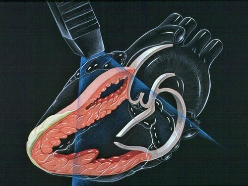
Fetal doppler ultrasound is a technique that uses high-frequency waves to investigate the movement, direction and velocity of blood flow in the blood vessels, heart, brain, and umbilical cord of the fetus. Thereby helping the doctor assess the health and development of the fetus and give appropriate treatment.
Compared with normal ultrasound, fetal doppler ultrasound has many outstanding advantages such as:
The procedure is not complicated. Appropriate cost. Safe, non-invasive, no harm to mother and fetus. High accuracy, the ability to detect problems earlier and more accurately.
2. Indication for doppler ultrasound 32 weeks pregnant
The obstetrician will appoint 32 weeks pregnant Doppler ultrasound to:
Assess the blood flow in parts of the fetus such as the umbilical cord, brain, heart to determine the ability of the fetus to absorb oxygen and nutrients . Investigation of blood flow through arterioles and veins. Thereby estimating fetal development. Screening for diseases and birth defects of the fetus, giving remedial measures and dealing with them as soon as possible. Assess the appropriate time of delivery.
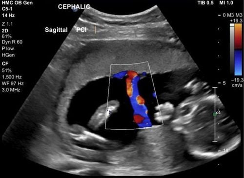
Siêu âm doppler thai 32 tuần giúp bác sĩ đánh giá tình trạng thai nhi
Subjects assigned to 32 weeks pregnant doppler ultrasound include:
Pregnant women with multiple pregnancies. The fetus is affected by Rh antibodies due to the mother's Rh incompatibility. The fetus is affected by Parvovirus disease - an infectious disease caused by the Canine Parvovirus virus. Pregnant women with a history of low birth weight babies. The fetus is growing slowly compared to the standard growth chart. Pregnant women with a history of late miscarriage or fetal death shortly after birth. Pregnant women with diseases during pregnancy such as hypertension, diabetes,... Pregnant women with low body mass index (BMI) or higher than standard. Pregnant women smoke during pregnancy. 32 weeks pregnant doppler ultrasound is an important time that women should not ignore because the last 3 months of pregnancy is when there is a risk of morphological abnormalities in blood vessels, heart, brain. This technique helps to assess the health status and development of the fetus comprehensively and detect birth defects and abnormalities, if any, early. Thereby, your doctor will develop a plan to ensure the pregnancy has the best outcome for both you and your baby.
Vinmec International General Hospital has full professional qualifications and technical means to effectively perform prenatal examination methods, including fetal Doppler ultrasound. There is a team of well-trained and experienced obstetricians, a complete and modern medical equipment system, professional service quality, effective diagnosis and treatment. contribute to the protection of pregnancy health for both mother and baby.
Please dial HOTLINE for more information or register for an appointment HERE. Download MyVinmec app to make appointments faster and to manage your bookings easily.