
Biological characteristics of T lymphocytes
Posted by MSc. Nguyen Van Office and MSc Le Thi Huyen - Cell Therapy Division, Vinmec High-Tech Center
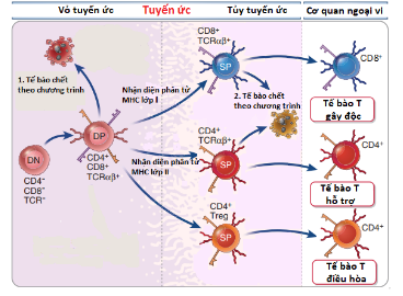
1. T-lymphocyte maturation in the thymus
T lymphocytes migrate from the bone marrow into the thymus to undergo "training" to become mature T-lymphocytes. In the thymus, the T-lymphocytes form surface receptors. T-cell receptor (TCR) cells enable them to recognize foreign antigens. Each TCR is a gene expression product from the arrangement of genes that regulate the T-cell receptor. These receptors then interact with thymic cortical epithelial cells to test their ability to recognize one of two types of MHC molecules (MHC class I and MHC class II). T lymphocytes that are able to recognize MHC class I molecules become CD8+ T cells, and T lymphocytes that are able to recognize MHC class II molecules become CD4+ T cells .
T cells continue to leave the thymus cortex and migrate into the thymus medulla to engage in cell recognition training. Here, T lymphocytes interact with myeloid epithelial cells that bind large numbers of the body's protein-presenting MHC molecules. During this process, T lymphocytes that are weakly bound to the MHC molecule are matured and directly enter the peripheral blood. In contrast, T cells with strong binding to MHC molecules will no longer be able to distinguish their own proteins from foreign agents, they will die by apoptosis to protect the body.
In our body hundreds of millions of T lymphocytes enter the thymus every day and only 2% to 4% of these cells become mature T lymphocytes. Therefore, the maturation process of T lymphocytes is always tightly controlled to ensure that they have the ability to recognize abnormal cells with their normal cells to destroy or activate other cells. exercise its immune function.
2. T lymphocyte classification
Based on the functional characteristics of the cells, T lymphocytes are classified into 5 main types of cells as follows:
Cytotoxic T cells (CD8+ T): Toxic T cells have the ability to recognize and destroy Abnormal cells such as virus-infected cells or cancer cells through specific binding of the T-cell receptor (TCR) with antigens presented on the MHC class I molecule of the abnormal cell.
Helper T cells ( CD4+ T ): Helper T cells are activated through specific binding of the T cell receptor (TCR) to foreign antigens presented on the surface of MHC class II molecules. of antigen-presenting cells.
Regulatory T cells: Regulatory T cells have a role in controlling the immune system and fighting autoimmune diseases.

Memory T cells: Memory T cells have a longer lifespan than other types of T cells and they have the ability to rapidly multiply to produce large numbers of effector T cells. upon re-exposure to antigens. Natural killer T cells (NKT): Natural killer T cells are a group of cells that have both T cell and natural killer (NK) characteristics. link between the innate and adaptive immune responses. Natural killer T cells, when activated, can perform the same function as cytotoxic and helper T cells.
REFERENCES
Abul K. Abbas, et al. (2017). T lymphocyte development. Cellular and Molecular Immunology, ninth edition, Elsevier, Philadelphia, 199-205. Bruce Alberts, et al. (2002). The Adaptive Immune System. Molecular Biology of the Cell , 4th edition, Garland Science, New York. Canoe RLE & Lopera HDE. (two thousand and thirteen). Introduction to T and B lymphocytes. Autoimmunity: From Bench to Bedside, El Rosario University Press; Bogota, chapter 5. Kurd N, Robey EA. T-cell selection in the thymus: a spatial and temporal perspective. Immunol Rev. 2016;271:114-126.
Dịch vụ từ Vinmec
-
 Các giai đoạn và cách điều trị ung thư bạch huyết tế bào B
Ung thư bạch huyết tế bào B đề cập đến một nhóm các bệnh ung thư tấn công hệ thống miễn dịch. Đây là loại ung thư hạch không Hodgkin phổ biến nhất, thường bắt đầu trong các hạch bạch ...
Đọc thêm -
 Tế bào giết tự nhiên và cơ chế hoạt động trong hệ miễn dịch bẩm sinh
Cơ chế điều hòa hoạt động của các tế bào NK thông qua các thụ thể hoạt hóa và ức chế là rất phức tạp. Bình thường, các thụ thể ức chế của tế bào NK liên kết với các ...
Đọc thêm -
 Tế bào giết tự nhiên (tế bào NK) – Vũ khí mạnh mẽ chống lại ung thư phổi tế bào nhỏ
Một loại tế bào miễn dịch được gọi là các tế bào giết tự nhiên (NK – Natural Killer) có khả năng trở thành một vũ khí mạnh mẽ trong cuộc chiến chống lại căn bệnh ung thư phổi, theo ...
Đọc thêm -
 Công dụng thuốc Blinatumomab
Bệnh nhân bạch cầu cấp đang chịu ảnh hưởng bởi tình trạng thần kinh có thể bắt đầu với thuốc Blinatumomab. Vậy thuốc Blinatumomab là thuốc gì, trong quá trình dùng thuốc có xảy ra tác dụng phụ hay không?
Đọc thêm -
 Sàng lọc hiệu năng cao các chất điều hòa dược học đối với cytokine điều hòa ức chế miễn dịch IL-10
Nghiên cứu khoa học về Sàng lọc hiệu năng cao các chất điều hòa dược học đối với cytokine điều hòa ức chế miễn dịch IL-10 được báo cáo tại Hội thảo Gen và tế bào Vinmec III diễn ra ...
Đọc thêm