
Atelectasis: Diagnosis and treatment
1. What is atelectasis?
Atelectasis can interfere with the airway, especially if there is additional lung disease. Treatment for this condition depends on the cause and severity of the atelectasis.
2. Diagnosis of atelectasis
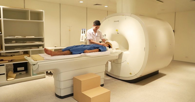
CT scan: Because CT is a more precise technique than X-ray, it can sometimes help better detect the cause and type of atelectasis. Oxygen measurement: This simple test uses a small device placed on your finger to measure the level of oxygen in your blood. It helps determine the severity of atelectasis. Thoracic ultrasound: This noninvasive test can help your doctor tell the difference between atelectasis, pulmonary sclerosis, pneumonia caused by fluid in the air sacs, and pleural effusion. Bronchoscopy: Using a flexible, lighted camera-tipped tube inserted down the throat allows the doctor to see what could be causing the blockage, such as a mucus plug, tumor, or foreign body. This method can also be used to remove blockages.
3. Treatment of atelectasis
3.1 Chest physiotherapy
Performing deep breathing exercises (spirometry) and using a device to assist with a deep cough can help clear secretions and increase lung volume. Position the body so that the head is lower than the chest (postural drainage). This allows the mucus to better drain from the bottom of the lungs. Tap the chest over the collapsed area to loosen the mucus. This technique is called the typing method. You can also use mechanical mucus cleaning devices, such as an air pulse vibrating vest or hand tools.
3.2 Surgery
If a tumor is causing atelectasis, treatment may include surgery to remove or shrink the tumor, with or without other cancer treatments (chemotherapy or radiation).

3.3 Breathing treatment
For children, childhood trauma is often caused by airway obstruction. To reduce the risk of illness, keep small objects out of the reach of children.
In adults, anemia often occurs after major surgery. If surgery is planned, talk to your specialist about strategies to reduce risk. Some research suggests that breathing exercises and muscle training may reduce the risk of disease after certain surgeries. In addition, smoking is also recommended because smoking increases mucus production and affects hair-like structures in the bronchial tubes (hairs).
Để đặt lịch khám tại viện, Quý khách vui lòng bấm số HOTLINE hoặc đặt lịch trực tiếp TẠI ĐÂY. Tải và đặt lịch khám tự động trên ứng dụng MyVinmec để quản lý, theo dõi lịch và đặt hẹn mọi lúc mọi nơi ngay trên ứng dụng.
Dịch vụ từ Vinmec
-
 Vì sao cần giải phóng đường thở trong cấp cứu?
Không chỉ với bệnh nhân bị dị vật đường thở mà cả những bệnh nhân bị ngất xỉu, ngừng thở, ngừng tuần hoàn thì đều cần phải giải phóng đường thở. Điều đó chứng tỏ việc khai thông đường thở ...
Đọc thêm -
 Công dụng thuốc Actidose
Actidose hay than hoạt tính là thuốc nằm trong nhóm thải độc. Tuy nhiên công dụng và độ an toàn của thuốc cần được kiểm tra kỹ lưỡng. Hy vọng những thông tin sau đây sẽ giúp bạn hiểu và ...
Đọc thêm -
 Người bị chấn thương sọ não đang hôn mê bị nghẽn tắc đường thở phải làm sao?
Bác sĩ cho em hỏi người bị chấn thương sọ não đang hôn mê bị nghẽn tắc đường thở phải làm sao? Em cảm ơn bác sĩ.
Đọc thêm -
 Công dụng thuốc Xopenex
Thuốc Xopenex công dụng trong điều trị hoặc dự phòng cho những người bị bệnh tắc nghẽn đường thở có thể hồi phục, bao gồm hen suyễn và một số vấn đề về hô hấp liên quan đến dị ứng. ...
Đọc thêm -
 Công dụng thuốc Prakuff
Thuốc Prakuff được bào chế dưới dạng siro, có công dụng trong điều trị ho do viêm nhiễm đường hô hấp. Tìm hiểu một số thông tin về công dụng, liều dùng và lưu ý khi sử dụng Prakuff sẽ ...
Đọc thêm