This is an automatically translated article.
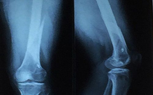
The article is expertly consulted by Master, Doctor Nguyen Hong Hai - Doctor of Radiology - Department of Diagnostic Imaging and Nuclear Medicine - Vinmec Times City International General Hospital.The knee joint is a rather loose joint, so during exercise or daily activities, it is easy to get injured. In order to diagnose the exact location of the injury at the knee joint quickly and effectively, doctors often assign patients to take x-rays of the knee joint.
1. When does the doctor order an X-ray of the knee joint?
The junction of the lower end of the femur, the upper end of the tibia, and the back of the kneecap is called the knee joint. In addition, around the knee joint, there is a system of ligaments around the joint, muscle cartilage and tendons...Doctors often assign patients to take X-rays of the knee joint in cases of injury caused by traffic accidents, accidents. work or accidents in daily life:
Dislocation of kneecap Fracture of cruciate ligament of knee Joint instability In addition, in some cases of following pathology, the doctor will also assign the patient to take x-ray. :
Knee Osteoarthritis Knee Osteoarthritis
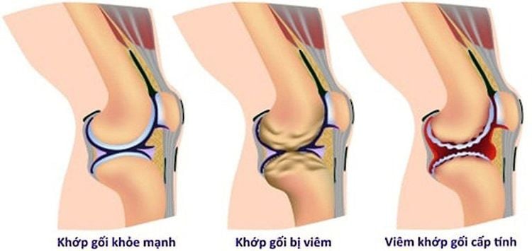
Bệnh nhân bị viêm khớp gối cũng được chỉ định chụp X-quang
2. X-ray procedure of straight knee joint
2.1 Preparation steps before taking x-ray of straight knee joint The patient receives an x-ray order from the doctor and moves to the designated X-ray location. Give the order form to the doctor in charge of taking X-rays and listen to instructions and explanations from the doctor. Expose the X-ray area as directed and remove jewelry if present near the X-ray unit. 2.2 Procedure for x-ray of straight knee joint The following are the steps to perform an x-ray of a rectilinear knee:2.2.1 Position of X-ray of a straight knee Instruct the patient to lie supine on the imaging table 2 arms extended, the leg on the right side of the foot should be rotated slightly into the back of the knee joint close to the center of the film vertically.
2.2.2 Position of X-ray of the lateral knee joint Instruct the patient to lie on the side to be imaged on the table, with the knee on the affected side flexed, the thigh slightly flexed, the lateral aspect of the thigh vertically aligned with the film. Adjust the convex shaft and pulley perpendicular to the film. The side leg does not need to be on the table and leans back as much as possible, the right hand needs to cover the headrest, the side hand does not need to grab the edge of the table.
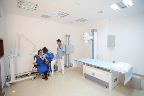
Bệnh nhân nằm trên bàn chụp x-quang và thao tác theo hướng dẫn của bác sĩ phụ trách
Get the knee joint in the middle of the film. The knee joint is clearly visible. The film has contrast sharpness, the film is clean without scratches. Have the patient's full name, P and T markings, date and year of the scan. Master, Doctor Nguyen Hong Hai graduated with a Master's degree in Diagnostic Imaging from Hanoi Medical University with strengths in diagnosing breast and thyroid cancer. Currently, Dr. Hai is working at the Department of Diagnostic Imaging, Vinmec Times City International Hospital.
Any questions that need to be answered by a specialist doctor as well as customers wishing to be examined and treated at Vinmec International General Hospital, you can contact Vinmec Health System nationwide or register online HERE.
MORE
Common problems in the knee joint The role of radiographs in diagnosing knee disease Diagnostic criteria for knee osteoarthritis


