This is an automatically translated article.
The article is professionally consulted by Master, Doctor Tong Diu Huong - Radiologist - Department of Diagnostic Imaging - Vinmec Nha Trang International General Hospital.X-ray of lobar pneumonia gives accurate results about the condition of the disease. Accordingly, the X-ray image of lobar pneumonia is a opacity that may occupy the entire lobe of the lung, usually the right lobe. However, it is necessary to distinguish lobar pneumonia from other diseases such as lobar tumor...
1. What is lobar pneumonia?
Lobar pneumonia is an acute type of pneumonia. X-ray of lobar pneumonia shows that the disease mainly affects the alveoli with large size, which may occupy the entire lung lobe or part of the lobe. The disease mainly occurs in the middle lobe of the right lung and is caused by pneumococci. The disease progresses through stages including: exudative, red hepatization and gray hepatization.

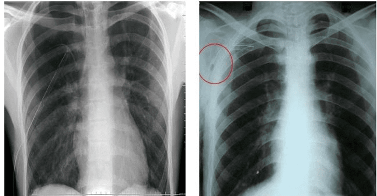
X quang viêm phổi thùy đóng vai trò quan trọng trong chẩn đoán phát hiện bệnh
2. X-ray image of lobar pneumonia
Currently, with the development of medical science, modern imaging techniques allow the assessment of lung lesions accurately. In particular, X-ray is still the first method to be performed in the initial disease diagnosis with accurate results.
The X-ray results of lobar pneumonia are as follows:
The pleura is blurred evenly. The lobar lesion was defined as a uniform and uniform opacification located in the lower half of the right lung. Lesions are large, triangular opacities that may occupy the entire right middle lobe, or the entire lobe of the lung. Based on X-ray images of lobar pneumonia to differentiate pneumonia from lobar pneumonia:
Lobar pneumonia: The lesion is a uniform opacities, occupying an entire lobe of the lung, usually the lower lobe of the lung. right lung. The blur has convex edges. Lung volume increases. Lobar lobar inflammation: The lesion is triangular opacities with convex edges, apex toward the hilum and base toward the periphery.
Trắc nghiệm: Làm thế nào để có một lá phổi khỏe mạnh?
Để nhận biết phổi của bạn có thật sự khỏe mạnh hay không và làm cách nào để có một lá phổi khỏe mạnh, bạn có thể thực hiện bài trắc nghiệm sau đây.3. X-ray of lobar pneumonia differentiated from atelectasis
X-ray images of lobar pneumonia allow differential diagnosis with atelectasis, specifically:
Lobar atelectasis shows a decrease in lobar or lobe lung volume, while lobar pneumonia increases the volume. . Lobar lesions are opacities with concave edges, while lobar pneumonia opacities are convex edges. Lobar collapse can cause pull on the adjacent lung. The X-ray image of lobar pneumonia is the basis for diagnosis, with the lesion being a uniform, triangular opacity. Imaging results play an important role in the differential diagnosis of whether the lesion is a partial lobe or the whole lobe of the lung, with damage from other pathologies such as atelectasis.
X-ray of lobar pneumonia gives accurate results about the condition of the disease, so that doctors will give the most accurate treatment regimen for the patient.

Hình ảnh X-quang cho phép chẩn đoán bệnh viêm phổi thùy
Currently, Vinmec International General Hospital is a general hospital that uses X-ray techniques as well as other modern techniques to diagnose and treat lung diseases. With modern facilities, quality medical services, a team of highly qualified and experienced doctors will bring optimal treatment results to customers.
To register for examination and treatment at Vinmec International General Hospital, you can contact Vinmec Health System nationwide, or register online HERE


