This is an automatically translated article.
This article was written by Specialist Doctor II Khong Tien Dat, Doctor of Radiology - Department of Diagnostic Imaging - Vinmec Ha Long International Hospital. The doctor has a lot of experience with more than 14 years working in the field of diagnostic imaging.When the pleural effusion is exuding, the pleural cavity is filled with fluid, causing pleural thickening with a clear image on the film. Therefore, X-ray is the most important laboratory test in the diagnosis of lung disease.
1. Why is there air in the pleura?
The pleura consists of parietal and visceral leaves, with a space between the two leaves. Normally in the void there is a little physiological fluid to lubricate when the lungs work to make the parietal and visceral leaves move rhythmically. When the amount of fluid exceeds the normal physiological index, there is a discharge that causes stagnation of fluid in the pleural cavity to a certain extent, causing pleural effusion.2. Causes of pleural thickening
There are many causes of air in the pleura, this can be a manifestation or complication of many different diseases such as:Pneumonia caused by bacteria of the cocci family, Hemophilus influenzae, St. pneumoniae, Mycoplasma pneumoniae, tuberculosis bacteria (Mycobacterium). Melanoma or lung cancer. Some diseases such as subdiaphragmatic abscess, liver abscess, cirrhosis ascites, pancreatitis, pericarditis, congestive heart failure. Trauma to the chest Chronic rheumatism or lupus erythematosus can also cause thickening of the pleura. Thickening of the pleura caused by parasites such as amoebic dysentery, filariasis, liver fluke disease.

3. Is the diagnosis of pneumothorax on X-ray accurate?
X-ray is the most important laboratory test in the diagnosis of lung disease. The value of radiographs in the diagnosis of lung disease is very high, only after the diagnosis of bone disease.When reading a chest X-ray, the first thing to do is to identify the underlying lung damage. Then it is necessary to determine the relationship of that lesion to surrounding organs.
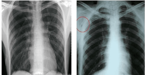
X-ray image of pneumothorax shows:
Complete opacity of a lung field. Solid opacity of the entire lung, pushing the organs involved. The intercostal space where the lesion is enlarged, the mediastinum is pushed to the healthy lung,... Homogeneous opacities at the hilum and the angle of the diaphragm, the upper boundary is not clear, forming the Damoiseau curve; The light image is usually peripheral and superior in the lung field, no pulmonary striations, limited to the lung parenchyma as the border of the visceral pleura. The lung parenchyma is pushed towards the hilum. In addition to chest X-ray, the careful analysis of this association will avoid confusion in the diagnosis of related diseases.

4. Treatment of pneumothorax
Treatment of primary spontaneous pneumothorax: The amount of pneumothorax is small, < 15% of the volume on the side of the pneumothorax; If the width of the air close to the pleura is < 2cm, then there is no need to aspirate, give the patient oxygen 2-3 liters / min for 2-3 days, then take a chest X-ray. Pneumothorax alone: Indicated for patients with primary spontaneous pneumothorax > 15% of the volume of the pneumothorax and the width of the proximal air band > 2 cm. Pneumothorax secondary to procedures: aspiration, pleural biopsy and transthoracic lung; If the volume is less than 15% of the volume in the pneumothorax, use a fine needle connected to the three prongs and a 50 ml syringe. After aspiration, the needle is withdrawn, if 4 liters of air is aspirated but the air is still out evenly, there is no feeling when it is exhausted, then pleural septal surgery is indicated. Pneumothorax is a fairly common medical condition today with many different causes. Based on each cause, the doctor will give the best emergency treatment plan for pneumothorax for the patient.Please dial HOTLINE for more information or register for an appointment HERE. Download MyVinmec app to make appointments faster and to manage your bookings easily.


