This is an automatically translated article.
The article is expertly consulted by Master, Doctor Nguyen Thi Mai Anh - Doctor of Radiology - Department of Diagnostic Imaging and Nuclear Medicine - Vinmec Times City International General Hospital.Magnetic resonance imaging (MRI) is a modern and effective subclinical imaging method, providing clear images for accurate diagnosis of breast lesions.
1. What is a mammogram?
Magnetic resonance imaging (MRI) is a method that uses a strong magnetic field, radio waves, and a computer to create detailed images of the internal structures of the breast.2. Why is an MRI of the breast needed?
Magnetic resonance mammography offers many benefits to both patients and physicians, including:It is used as an additional tool for breast screening after mammography and ultrasound or can be used in screening women at high risk for breast cancer Assess the extent of cancer after diagnosis or further evaluate for abnormalities on mammograms. Safe because no X-ray is used, the patient is not affected by ionizing radiation. Is the best method to assess the condition of cosmetic breast implants. Besides the benefits, MRI of the breast still has certain limitations. Breast MRI is not a substitute for mammography. Although this is a highly effective test, sometimes a mammogram may not find cancer. MRI of the breast can also lead to false-positive results. This means that the test finds a mass or other change, but it is not the actual cancer. If this happens, your doctor may recommend an ultrasound. If the suspected cancer is still not detected by ultrasound, the doctor will ask the patient to do an MRI biopsy.

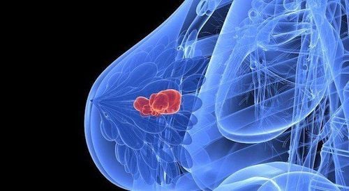
Chụp MRI giúp sàng lọc ung thư vú ở những phụ nữ có nguy cơ mắc bệnh cao
3. Indications for mammography
Below are the cases in which breast MRI is indicated, including:In cases where it cannot be concluded by conventional mammograms, it is indicated to use breast MRI to determine the breast lesions. love. Provide assessments of the extent of spread of breast cancer lesions after diagnosis. Examination of the contralateral breast for patients with breast cancer breast cancer. Evaluation of deep cancer invasion to the fascia. Evaluation of residual lesions after tumor resection. Suspect tumor recurrence in patients with or without postoperative mammography. Assess before, during, and after chemotherapy. Breast cancer screening in high-risk patients. There is malignant axillary lymph node involvement in which the primary tumor has not been detected. Investigate cases of very large breasts with or without breast implants. Evaluation of the integrity of cosmetic breast implants.
4. Preparing for a breast MRI
For best results, you should schedule your exam at certain times of your menstrual cycle. For example, if you're perimenopausal, the MRI facility may ask you to schedule your procedure between 5 and 15 days of your menstrual cycle. You should let them know what day of your period you are on so that the facility can arrange the best time for your breast MRI.Once you have scheduled your breast MRI, you will receive detailed instructions on how to prepare. Here are some general suggestions:
Tell your doctor about all the medicines you are taking, as well as any drug allergies or other medical conditions you have. Tell your doctor if you are pregnant. Women who are breastfeeding should also inform their doctor. Breast MRI conducted while a woman is breastfeeding may not produce images that are clear enough for accurate interpretation. Doctors recommend that you stop breastfeeding 2 days after having an MRI with contrast injection to give your body time to get rid of the drug. Before having a breast MRI, you should pump and store milk to feed your baby. Tell your doctor if you have any implanted medical devices. These can cause serious complications when exposed to strong magnetic MRI. For example, pacemaker, defibrillator, or implant, etc. Absolutely do not bring anything metal during breast MRI.

Hãy cho bác sĩ biết về tất cả các loại thuốc bạn đang dùng trước khi chụp MRI tuyến vú
5. Before breast MRI
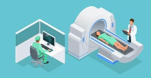
Once you have been assigned a breast MRI, you will move to the imaging department. Here, medical staff will welcome and guide you to change and remove jewelry and metal objects you bring. When you are done changing, you will be placed on the bed in a comfortable position suitable for the part to be photographed; then the bed will automatically move to the shooting area.In case you have claustrophobia, tell your doctor to be given a light sedative before the scan.
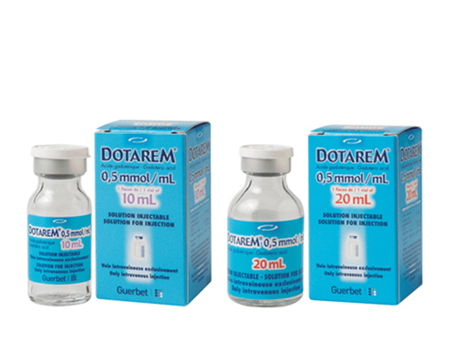
Next, the doctor will inject a magnetic contrast agent into a vein in your arm, to make the tissue or blood vessels visible on the MRI. The injection time is about 1-2 minutes. When the medicine is injected, you may feel a warm feeling all over your body, or your tongue may taste bitter. These symptoms should go away within 2-5 minutes.
6. During a breast MRI
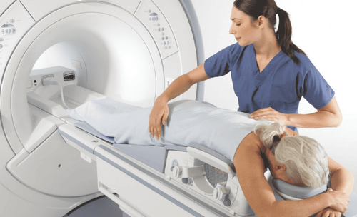
Your doctor will help position you on a padded table specifically designed for breast MRI. You will lie face down, arms by your side, and your head resting on a pillow. Your breasts are housed in two holes on an inner table that contains coils, which detect magnetic signals from the MRI machine. There is a large round hole in the center of the breast MRI machine, the entire table will then slide inside this round hole of the machine.You will need to lie still throughout 2 to 6 image sequences. Each sequence will last up to 15 minutes. When shooting, the camera will emit a loud sound due to magnetic resonance. In this case, you can wear headphones to relax, help reduce noise.
7. After breast MRI

Bệnh viện Đa khoa Quốc tế Vinmec là đơn vị đầu tiên đưa vào sử dụng máy chụp cộng hưởng từ 3.0 Tesla công nghệ Silent, mang đến những ưu điểm vượt trội
Vinmec International General Hospital is the first unit to put into use a 3.0 Tesla silent magnetic resonance imaging machine, bringing outstanding advantages. Magnetic resonance imaging (MRI) at Vinmec will have the following outstanding advantages:
High imaging technology, first-class safety by accuracy, non-invasiveness and no radiation. High image quality, allowing the doctor to make a comprehensive assessment, limiting the omission of injuries in organs, minimizing noise, creating the most comfort for the patient when taking pictures, reducing stress, this help better image quality and shorten the maximum scanning time. MRI at Vinmec can take 3D vascular reconstruction without injection of magnetic contrast, can reconstruct and handle motion disturbances of the patient Master, Doctor Nguyen Thi Mai Anh has had nearly 10 years of experience in the field of diagnostic imaging, especially in diagnosing breast and thyroid cancer. Received formal training at Thai Binh Medical University and specialized postgraduate training at Hanoi Medical University. Currently, the doctor is a radiologist at Vinmec Times City International General Hospital.
Please dial HOTLINE for more information or register for an appointment HERE. Download MyVinmec app to make appointments faster and to manage your bookings easily.
Reference source: Cancer.net


