This is an automatically translated article.
The article is professionally consulted by Master, Doctor Le Xuan Thiep - Radiologist - Department of Diagnostic Imaging - Vinmec Ha Long International Hospital. The doctor has extensive experience in the field of diagnostic imaging.Currently, magnetic resonance imaging (MRI) is increasingly popular because of its advantages in diagnosing diseases. Thanks to magnetic resonance, doctors will have more accurate image diagnosis than other paraclinical methods to survey a variety of parts of the patient's body.
1. What is resonance imaging (MRI)?
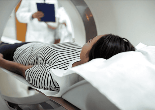
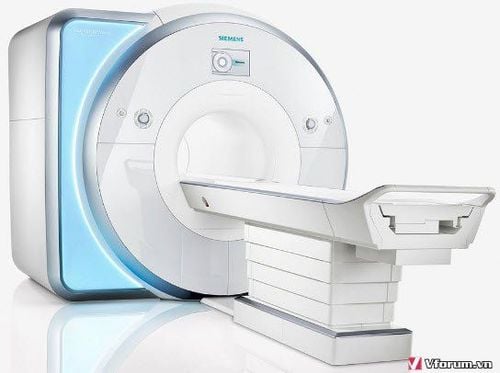
Magnetic Resonance Imaging (MRI) is a non-invasive imaging technique that uses a magnetic field and radio waves to help examine organs, tissues, and bones inside the body. When the body enters the magnetic resonance imaging process, magnetic waves and radio waves will affect the hydrogen atoms in the body, causing these atoms to absorb and release RF energy. The machine will then receive, process and convert the signals received during RF release into images displayed on the screen.MRI images with high quality and contrast, detailed, sharp, clear, have the ability to reproduce 3D anatomy well to help accurately diagnose the patient's pathology easily. than. In addition, using MRI without radiation, ensuring high safety, should be highly evaluated by medical experts.
2. Which part of the body is scanned for magnetic resonance imaging?

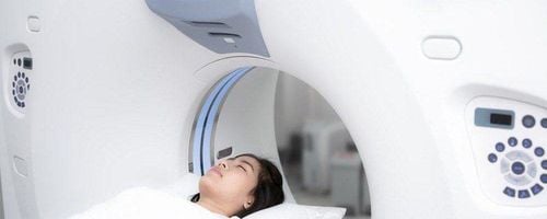
Đa số các bộ phận trên cơ thể đều có thể được khảo sát bằng MRI
Brain aneurysm Disease Eye and Inner Ear Treatment Multiple Sclerosis Spinal cord disease Stroke Brain injury Types of neuromas Cerebral vascular malformations Epilepsy In addition, special techniques Functional MRI of the brain (fMRI) also helps to create Visualize blood flow to certain areas of the brain, helping to determine which parts of the brain are handling important functions. From there, the doctor will identify the important areas that control movement and language in the brain for people who are assigned to have brain surgery or evaluate damage from head trauma, Alzheimer's disease, Parkinson's disease ... Orbital MRI can also help evaluate visual pathologies or lesions affecting the optic nerve, orbital tumors, cavernous carotid artery disease (causing protrusion).
An MRI of the heart and blood vessels helps to investigate some of the following:
Size and function of the heart chambers Thickness and movement of the walls of the heart chambers The extent of damage caused by a heart attack or heart disease. Structural problems of the aorta such as aneurysms or dissections Inflammation or blockages in blood vessels Evaluation of myocardial function, myocardial necrosis, myocardial integrity... consequences of ischemic disease - infarction Myocardial bleeding... With modern machines, it is even possible to evaluate and diagnose coronary artery disease (this is the strength of MSCT because of its ability to quickly capture and avoid image noise) MRI scans of internal organs Organs such as liver, gallbladder, kidney, spleen, pancreas, uterus-ovarian, prostate gland help detect tumors or other abnormalities. Meanwhile, bone and joint MRI will help evaluate the following areas:
Spine: spondylolisthesis, hernia, discitis or spinal cord disease such as trauma, spinal tumor Cartilage, structural diseases joints, tendons, bones and ligaments,... Especially in the pathologies of injury and injury to the ligaments, arthritis... MRI is considered an absolute strength compared to other diagnostic methods. Other images. Bone infections Tumors in bones and soft tissues For women, a breast MRI combined with a mammogram is used to detect breast cancer, especially in women with dense breast tissue or those with people at high risk of this cancer, monitor for recurrence after treatment, evaluate tumor lesions, lymph node metastases, chest wall metastases ... before conducting surgery or neo-chemotherapy.
3. Some notes when taking magnetic resonance imaging
Patients should note, MRI uses magnets with strong magnetic fields, so the presence of metal in the body is a threat if magnets attract or alter the MRI image. Therefore, before taking an MRI, the patient needs to perform a survey about the metal or electronic implanted devices in the body. Except for the cases where the device is certified to be safe with MRI, all the following implanted devices are not indicated for magnetic resonance imaging:Metal prosthetic joint Artificial heart valve Defibrillator Syringe Implants Pacemaker Pacemakers Metal clips, staples, clips in surgery Metal pieces Cochlear implants Some intrauterine devices MRI has no special requirements for eating but when conducting the patient may be asked to change into a gown and remove items that can affect magnetism such as jewelry, hairpins, watches, eyeglasses, wigs, dentures,...

Trước khi chụp Cộng hưởng từ, bệnh nhân sẽ được yêu cầu tháo bỏ trang sức, kính mắt,... những thứ có thể ảnh hưởng đến từ tính
Silent technology is especially beneficial for patients who are children, the elderly, weak health patients and patients undergoing surgery. Limiting noise, creating comfort and reducing stress for customers during the shooting process, helping to capture better quality images and shorten the shooting time. Magnetic resonance imaging technology is the technology applied in the most popular and safest imaging method today because of its accuracy, non-invasiveness and non-X-ray use.
Please dial HOTLINE for more information or register for an appointment HERE. Download MyVinmec app to make appointments faster and to manage your bookings easily.
MOREDoes Magnetic Resonance Imaging (MRI) require fasting? What is the effect of magnetic resonance imaging (MRI) of the brain? For the first time in Southeast Asia, Vinmec uses a magnetic resonance imaging machine with Silent technology


