This is an automatically translated article.
The article is professionally consulted by Doctor Nguyen Thi Le - Department of Obstetrics and Gynecology - Vinmec International Hospital Da Nang.Most children are born healthy, but there are still about 2-3% of fetuses with birth defects, causing severe physical and mental consequences for children. These birth defects can mostly be detected through ultrasound screening for fetal malformations during pregnancy. Let's learn about this ultrasound method through the following article!
1. Common types of fetal malformations
Screening for fetal malformations during pregnancy can detect and intervene early in the fetus or treat early after birth, helping children develop normally. In addition, this also helps parents make decisions to keep or abort the fetus if the fetus has too severe defects, it is difficult to survive or develop after being born.Common birth defects
● Down syndrome, Edwards syndrome
Growth retardation, mental retardation
Sex disorder, inability to develop sex
● Cleft lip, cleft palate
● Congenital heart, hypothyroidism
Defects in the nervous system, head, face, neck, chest, abdomen..
Deformities of extremities, malformations of genitals
Trắc nghiệm: Xét nghiệm Triple test là gì? Cần thực hiện khi nào?
Triple test và một trong những xét nghiệm sàng lọc trước sinh quan trọng nhất trong thai kỳ, giúp chẩn đoán nguy cơ dị tật thai nhi, là cơ sở để các bác sĩ chỉ định thực hiện các xét nghiệm sàng lọc xâm lấn như chọc ối, sinh thiết gai nhau. Theo dõi bài viết sau để biết Triple test là gì và nên thực hiện khi nào?2. Ultrasound screening for fetal anomalies
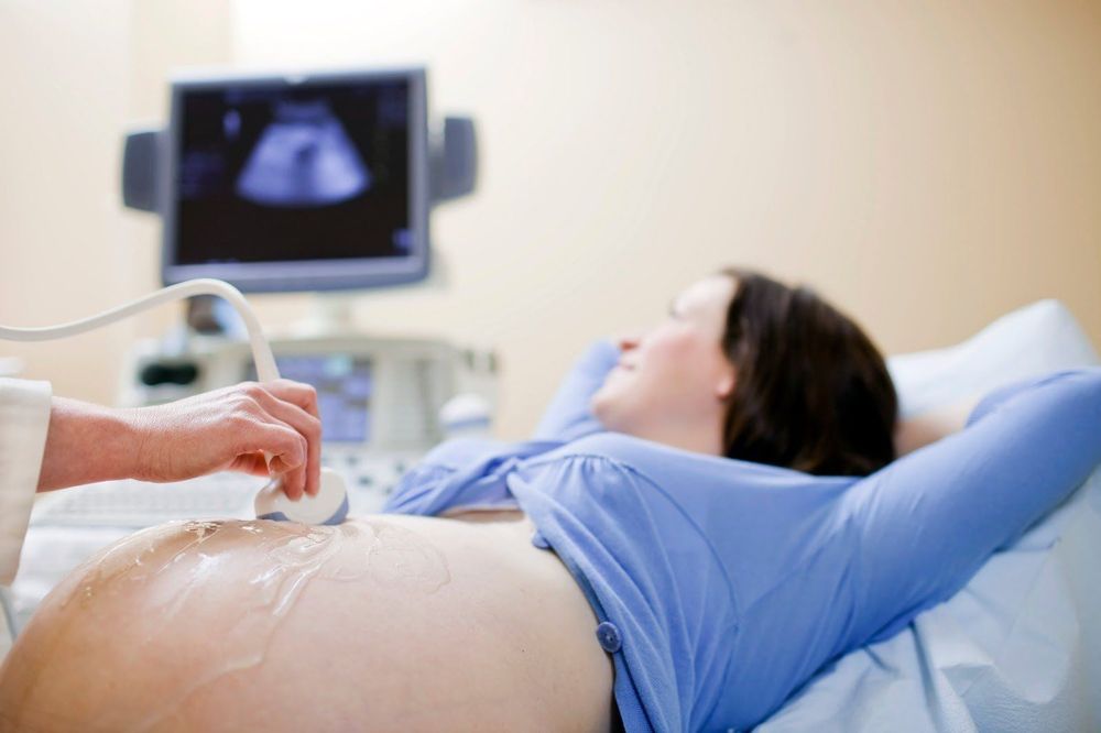
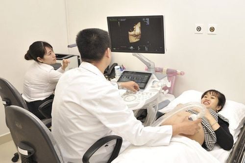
Fetal ultrasound uses ultrasound waves to image and observe the structure and functioning of the fetus in the uterus. Ultrasound is painless, has no side effects, and can be performed at any time during pregnancy. Depending on the specific case, the doctor may specify the time and number of ultrasounds for each person differently.2.1. Is pregnancy ultrasound safe? Ultrasound has been used in obstetrics and gynecology for decades and has a complete set of standard and procedure guidelines. Up to now, no scientists have confirmed that ultrasound is harmful to the health of mothers and babies (according to research from the British Medical Ultrasound Society - Guidelines For The Safe Use of Diagnostic Ultrasound Equipment). However, the recommendation of ISOUG (World Society of Obstetrics and Gynecology) is that the duration of fetal ultrasound should be minimized, using the shortest time and lowest wave intensity possible to obtain sufficient information. for diagnosis.
2.2. How long does an ultrasound scan for fetal malformations take? Normally, ultrasound screening for fetal malformations takes about 15-30 minutes. However, with some exceptions it may take longer. For example, when the pregnancy is difficult to assess due to the difficult position; a lot of movement, or when the mother is overweight; If the abdominal wall tissue is thick, preventing ultrasound waves from passing, it will be difficult for the doctor to get a good view to check and evaluate the fetal condition. There are even cases where the doctor has to make an appointment for the next time.

3. Why is it necessary to have an ultrasound to screen for fetal malformations?
With the current momentum of modern science and technology, many birth defects can be successfully treated if detected early by prenatal screening and diagnosis. This can help detect birth defects early as well as provide timely intervention measures, thereby minimizing birth defects in the womb and thus, babies born can develop normally.You should perform prenatal screening by periodic antenatal check-ups, fetal malformation ultrasound screening and testing during pregnancy. With these prenatal diagnostic tests, couples will know there is an 80-90% chance that their baby is healthy or has any problems.
4. Cases requiring prenatal diagnosis
In some cases, prenatal diagnostic tests should be done to screen and detect birth defects of the fetus early, including:
● Pregnant women over 35 years old
Inbreeding
● Pregnant women with a family history of birth defects
● Pregnant women with a history of premature birth, miscarriage, or stillbirth of unknown cause
● Pregnant women infected in the first 3 months of pregnancy: rubella, colds, flu, medical illnesses...
● Parents often have to work and live in toxic and chemical-laden environments
For normal, low-risk healthy women, it is still recommended to perform ultrasound negative screening for malformations at important milestones: 12 weeks, 22 weeks and 32 weeks gestation. However, cases with high risk or suspected abnormality can be screened for malformations by ultrasound as prescribed by the antenatal clinician.
5. Timing of ultrasound screening for fetal malformations

5.1. From 11 weeks - 13 weeks 6 days This time, the doctor will determine the most accurate gestational age. The doctor can also check how the fetus is, whether the fetus is single or multiple, or whether the fetus is developing normally or not?
Besides, the doctor can detect some abnormalities such as: anencephaly, umbilical hernia, open abdominal wall...; survey some signs suggestive of fetal chromosomal disorders: nasal bones, nuchal translucency, vascular abnormalities under doppler ultrasound... Simultaneously combined with maternal blood tests to determine the risk children with Down syndrome, pre-eclampsia-eclampsia, fetal growth retardation in the uterus.
Based on high or low screening risk, you may be advised to have a diagnostic test for fetal pathology.
This first-trimester ultrasound also examines the mother's gynecological abnormalities inside and outside the uterus.
5.2. From 18-22 weeks This is an important period for doctors to survey fetal morphology, detect birth defects, cervical length to assess the risk of preterm birth.
With fetal abnormalities, you can be consulted with amniocentesis under ultrasound to support the diagnosis of fetal disease.
Note, the termination of pregnancy can only be done before the 28th week. After this time, if premature birth is stimulated, the malformed fetus can still live, so the prenatal diagnosis will no longer be meaningful. means.
5.3. From 30 to 32 weeks An ultrasound of the fetus during this time is more likely to detect more abnormalities that appear late, appear during fetal development such as abnormalities in the arteries, in the heart or in one of the blood vessels. structural regions of the brain. In addition, ultrasound helps to assess fetal health, amniotic fluid status and prognosis for delivery.
6. How to prevent birth defects
There are cases of fetal malformations stemming from improper nutrition during pregnancy and environmental factors. In addition, women should prepare pre-pregnancy knowledge that will help prevent birth defects6.1. Before becoming pregnant When you intend to become pregnant, you should have a general health examination to detect and definitively treat dangerous diseases that can affect the fetus. Specifically, you should be vaccinated against diseases such as flu, rubella, hepatitis B...at least 3 months before becoming pregnant. In addition, women who are planning to become pregnant should start taking iron supplements and especially folic acid, to avoid the risk of neural tube defects in the fetus.
6.2. During pregnancy Pregnant women can limit the chance of birth defects in the following ways:
Prenatal check-up under the guidance of a doctor
● Do not drink alcohol, smoke
● Notice Talk to your doctor about diseases that run in the family
Take adequate vitamins and minerals every day
Use medications with caution
Maintain a healthy lifestyle and maintain an ideal weight
Carry out vaccinations against common diseases
Periodic antenatal check-up, complete screening tests
After planning to have a baby, you should prepare a small notebook to store necessary information, for example such as testing for fetal malformations at what week, during pregnancy, what should be added and what foods should be avoided... This can help you limit the dangers during pregnancy.
With non-invasive prenatal screening (NIPT) based on fetal DNA testing in mother's blood, doctors can detect fetal abnormalities as early as 9 weeks old instead of waiting until 12 weeks of age as before, while also reducing the risk of miscarriage compared with conventional amniocentesis. This advanced technique is being implemented at Vinmec International General Hospital, bringing outstanding improvements in the results of prenatal screening, helping to intervene early and effectively with fetuses with anomalies. Non-invasive prenatal screening method (NIPT) is considered the "key" to "decode" fetal malformations from a very young gestational age.
In addition, to protect mother and baby during pregnancy, Vinmec provides a package Maternity service to help monitor the health status of mother and baby comprehensively, periodically check antenatal care with leading obstetricians and gynecologists. , perform a full range of tests, important screening for pregnant women, timely advice and intervention when detecting abnormalities in the health of mother and baby.
Please dial HOTLINE for more information or register for an appointment HERE. Download MyVinmec app to make appointments faster and to manage your bookings easily.


