This is an automatically translated article.
DSA is a diagnostic imaging method that combines X-ray and digital processing. When taking a DSA, the patient must insert instruments in the groin, at that location there may be bleeding or hematoma. Therefore, for a DSA, the patient will have to lie motionless for 24 hours.
1. What is a DSA scan?
1.1 What is DSA?DSA stands for Digital Subtrasion Angiography, is an imaging method combining X-ray and digital processing that uses an algorithm to remove the background on 2 images obtained before and after injection. Contrast into the patient's body, for the purpose of studying blood vessels in the body.
A DSA scan will obtain images that provide accurate information about the blood circulation as it passes through all parts of the body, especially the blood supply to the brain and heart. From there, the doctor can soon detect abnormal conditions of blood vessels such as: narrowed vessels, aneurysms, blockages, ... or some diseases such as myocardial infarction, vascular accident. This helps in the accurate diagnosis and treatment of serious diseases related to the blood circulation in the body, planning the anatomy helps to pinpoint the exact location of the damage to the side. in the body.
1.2 DSA system and working mechanism
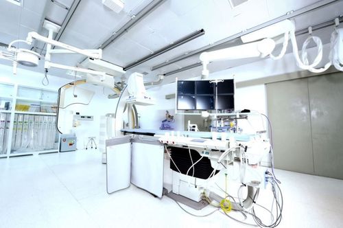
A DSA system usually includes components such as:
X-ray generator, image acquisition unit, digital image processing unit, display unit,... The central part of the DSA system is the digital image processor. It will collect images from video camera and generate pulse control signal in time for both X-ray generator and image acquisition system in order to control the signal reception and transmission between the two systems. this. The image acquisition process is started when the shock over time is transmitted to the radiating unit, then X-rays are emitted, pass through the patient's body and are received by the optical tube. A lens is placed between the optical tube and the video camera to limit the amount of light that reaches the camera.

Chụp DSA sẽ thu được những hình ảnh cung cấp thông tin chính xác về quá trình lưu thông máu
After receiving the light signal from the optical tube, the video camera converts to video signals and feeds them to the image processor as analog. At the image processing unit, the analog signal will be digitized and stored in memory, which is used to compare and remove the background with another image at a different time or at a different energy level. .
A common algorithm in digital X-ray systems used for background removal is a time-based erasure algorithm. During the implementation of this algorithm, the original image is acquired. The contrast material is then given into the body through a vein. Next is the process of acquiring an animated image as the contrast material enters the body in a preset time unit. At this time, the image processing unit will take the image obtained when there is no contrast agent as the background image and proceed to exclude the background image with the image obtained when there is contrast for the same static anatomical structures. between two images.
2. In what cases is DSA diagnosed?
DSA imaging technique is often used for diagnosis and treatment in some cases such as:
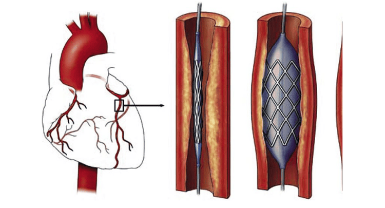
Diagnostic and interventional coronary artery bypass surgery Uterine artery embolization to treat uterine fibroids, uterine bleeding Bronchial artery occlusion for the treatment of hemoptysis Petrochemical embolization for the treatment of liver cancer Placement of artery stents Femoral angiography Diagnostic and interventional treatment of carotid and cerebral artery diseases Placing a generator heart rate Bronchial and uterine artery embolization Uterine fibroids, liver tumors Peripheral angiography

Kỹ thuật chụp DSA thường được dùng để chẩn đoán và can thiệp động mạch vành
3. Can a DSA scan show movement?
Before conducting a DSA scan, the patient will be injected by doctors with a dye, also known as a contrast agent, which is responsible for brightening blood vessels. Contrast is completely harmless to the body and will be excreted in the urine. The DSA scan process is painless, at first after the injection, the patient may feel a little uncomfortable. During the DSA scan, the patient will be cared for by medical staff, and the pulse and heart rate will be monitored during the DSA scan. The camera rotates 360 degrees and captures a complex series of images.
When taking a DSA, the doctor will thread the instruments in the groin, after doing so, there may be bleeding or hematoma, so the doctor will conduct a compression bandage in the groin. The patient's leg will now be immobilized for 8 hours, and the patient will remain motionless for 24 hours until the doctor removes the compression bandage. Therefore, after taking DSA, patients need to be supported by relatives in daily activities such as eating, urinating,...
In short, DSA is an imaging method that combines X-rays. - Optical and digital processing, helping doctors to detect abnormal conditions of blood vessels early. Before the scan, the patient will have to inject contrast dye to brighten the blood vessels. In addition, during a DSA, the patient has to insert instruments in the groin, at that location there may be bleeding or hematoma, so the doctor will apply compression in the groin. Therefore, for a DSA, the patient will have to lie motionless for 24 hours until the doctor removes the compression bandage.
Vinmec International General Hospital with a system of modern facilities, medical equipment and a team of experts and doctors with many years of experience in medical examination and treatment, patients can rest assured to visit. examination and treatment at the Hospital.
Please dial HOTLINE for more information or register for an appointment HERE. Download MyVinmec app to make appointments faster and to manage your bookings easily.
SEE MORE
Background erasing angiography and pulmonary vascular intervention Advantages of DSA background angiography Which cases need magnetic resonance imaging (MRI) of the brain?


