This is an automatically translated article.
The article was professionally consulted by Specialist Doctor I Pham Thi Yen - Obstetrics and Gynecology Department - Vinmec Hai Phong International General HospitalCesarean section is an intervention to remove the fetus and its appendages from the uterus through the abdominal wall incision and the uterine incision. Indication for cesarean section during labor is not safe for the mother and fetus, in cases where labor is forced but cannot induce labor, when delivery is difficult or because the characteristics of the fetus make it impossible to give birth. and in an emergency situation requiring a quick removal of the fetus, but giving birth below is not eligible.
1. Indication for cesarean section
1.1. Indications for prophylactic cesarean section (active) Abnormal pelvis
Totally narrow pelvis: all diameters of the pelvis are reduced including upper and lower waist. Especially, defender protrusion diameter from 8.5 cm or less Distorted pelvis (displaced pelvis or asymmetrical pelvis): measured Michaelis fillings are not symmetrical and diameter of protrusion-defender is measured from 8.5cm or more down. Funnel-shaped pelvis: the pelvis is deformed, making the lower waist narrow, and the upper waist wide. The pregnancy can easily pass through the upper waist, but it is difficult or impossible to pass through the lower waist. The pelvis is measured by the diameter of the biceps. If this diameter is small, about 9cm, the fetus will not be delivered, so an elective cesarean section is indicated. In the above cases, it is necessary to do more test of the crown to challenge the lower birth canal if the pelvis is limited (the fetus is not large), if it fails, there is an indication for cesarean section.
Fetal outlet is obstructed
Tumor of the prostate: ovarian tumor located deep in the pelvis, most commonly fibroids in the isthmus or cervix, ovarian cyst, other tumors located in the path pregnancy comes out. Some rare prostate tumors are vaginal tumor, fallopian tube tumor, wide ligament tumor, pelvic tumor such as kidney tumor, rectal tumor, bladder tumor, double uterus Central placenta previa or placenta The striker caused a lot of bleeding, forcing emergency surgery to stop the bleeding to save the mother. In the case of central placenta previa, it is necessary to rely on ultrasound results to make an indication for elective cesarean section. Uterus has a scar from surgery in the following case
Scars of previous surgery on the body of the uterus: scar removal fibroids, scars of hysteroplasty, suture scars of rupture, uterine perforation, scars of corner-cutting surgery, uterine horns. Scars from two or more transverse cesarean sections of the uterus or previous cesarean section less than 24 months ago.
Indications for caesarean section for the mother's cause
The mother has chronic or acute systemic diseases, if the lower birth canal may pose a risk to the mother's life (severe heart disease, hypertension, severe preeclampsia and eclampsia). Abnormalities in the lower genital tract of the mother such as: vaginal stricture (congenital or acquired), history of fistula, caesarean section. Abnormalities of the uterus such as: double uterus (womb that is not pregnant often becomes tumor of the prostate), bicornuate uterus... especially when accompanied by abnormal fetal position. Causes on the side of the fetus
The fetus is malnourished/severe intrauterine growth retardation The fetus has a blood type disagreement with the mother, if the fetus is not removed, there is a possibility that the fetus will still be stillborn in the uterus

1.2. Indication for caesarean section during labor Indication for caesarean section for the mother's cause
Older child is a pregnant woman aged 35 or older. May or may not include reasons for infertility: history of infertility treatment, rare children. Maternal medical conditions that may still permit monitoring of labor will be performed by cesarean section if another factor of difficulty is present. Indications for caesarean section for the cause of the fetus
Larger than 4,000g fetus is not due to abnormal pregnancy. Abnormal positions: shoulder/horizontal, forehead, fontanel, posterior chin, buttocks. Multiple pregnancy: if the first pregnancy is not the first. Labor has progressed to fetal failure when there are not enough conditions for lower birth. Indications for cesarean section because of abnormalities in labor
Abnormal uterine contractions after using drugs to increase or decrease contractions to correct without success. The cervix is not dilated or dilated although the uterine muscles are synchronous, consistent with the cervical dilation. Premature/premature rupture of membranes causes labor to stop progressing and induce labor to fail. Birth disproportionate to the pelvis. The head does not enter when the cervix is fully dilated, although the uterine contractions are strong enough, it is possible that due to asymmetry, the pelvis is quite discreet and unknown. 1.3. Indications for cesarean section for complications in labor Bleeding because of placenta previa, placental abruption. Threatened rupture and rupture of the uterus. Umbilical cord prolapse while the fetus is still alive. Sa chi after trying to push up but failed.
2. C-section technique
Step 1. Anesthesia:
General anesthesia or epidural or spinal anesthesia to ensure that the mother does not wake up during the cesarean section.
Step 2. Intra-abdominal:
Incision below the midline below the navel or the horizontal line on the abdomen depending on the surgeon's ability, the state of the mother and the fetus Incision of the subcutaneous fat layer Incision of the white line between the two rectus muscles (with You can make a small incision and then separate it with your fingers) if you have a cesarean section along the longitudinal line. With the horizontal cesarean section, the fascia is made on both sides according to that incision, separating the fascia from the broad muscle layer and then opening. line between the two rectus abdominis. The surgeon pairs the peritoneum with a toothless surgical forceps, and the contralateral peritoneum clamps the opposite side with a toothless hemostatic forceps. The surgeon and the surgical assistant took turns releasing and re-pairing, then using a knife or scissors to open a hole in the peritoneum. Use scissors to extend the peritoneum above and below. Insert wet gauze on both sides, leaving the cord out Place the valve over the bladder guard and expose the lower part of the uterus Incision of the peritoneum horizontally about 2cm below the “tight line of the peritoneum” Use curved scissors to separate the peritoneum The fascia can be removed from the lower segment up and down, the nose is curved upwards to avoid injury to the uterine artery. Using a knife, make a small incision 1-2 cm across the lower segment, then use 2 index fingers to tear the incision to the left. 2 sides

Step 3. Remove the fetus and placenta from the uterus:
The surgeon removes the fetus with her left hand while the woman sucks blood and amniotic fluid (if the head is too high, forceps can be used) After the crown is exposed incision, the assistant presses the bottom of the uterus to help the baby's head come out. In the case of transverse position: take the fetus by the feet of the fetus. In case of breech position: take the pregnancy by the buttocks (breast breech position) or by foot (complete breech position) Wipe dry, clamp the umbilical cord slowly, remove the fetus to wipe clean, lay the baby right on the mother's chest (if the mother is able to) spinal or epidural anaesthesia) Inject 10 units of oxytocin into a flowing infusion bottle and allow it to flow rapidly for a good contraction of the uterus (do not inject oxytocin directly into the vein) Perform vegetable extraction, clean the uterine cavity with gauze. If at cesarean section, the woman is not in labor, dilate the cervix with a finger and then change the cast iron. Step 4. Sewing to restore the uterine muscle:
Restoring the lower uterine muscle with Vicryl 0 thread, starting with stitching the 2 corners of the uterus, avoiding missing corners Continue suturing the myometrium with continuous or separate stitches 1cm apart. , can add a 2nd layer to bury the first layer, check hemostasis Cover the peritoneal cavity with Catgut 00 suture with continuous stitches, check hemostasis Remove valve on the guard, take gauze, clean the abdomen, check Examine the 2 ovaries, 2 fallopian tubes, posterior uterine surface and posterior sac with Douglas Step 5. Abdominal closure:
Sew the abdominal wall peritoneum with Catgut 00 suture with continuous stitches Sew the 2 rectus abdominis close together. with 2-3 stitches Catgut stitches separate stitches Sew with Vicryl thread balance 0. If the fat layer is thick, sew with Catgut stitches with separate stitches or continuous stitches Sew the skin with linen thread, loose stitches or continuous stitches under the skin with small Vicryl threads Antiseptic Close the incision and a sterile bandage The surgeon keeps his hands clean to take blood from the vagina and see if the uterus is shrinking well, disinfect the vagina Wipe the blood off the patient before moving into the room recovery. The cesarean section usually takes about 30-40 minutes. It only takes 5 minutes after the mother's belly incision until the baby is out. The rest of the time is suturing the wound.
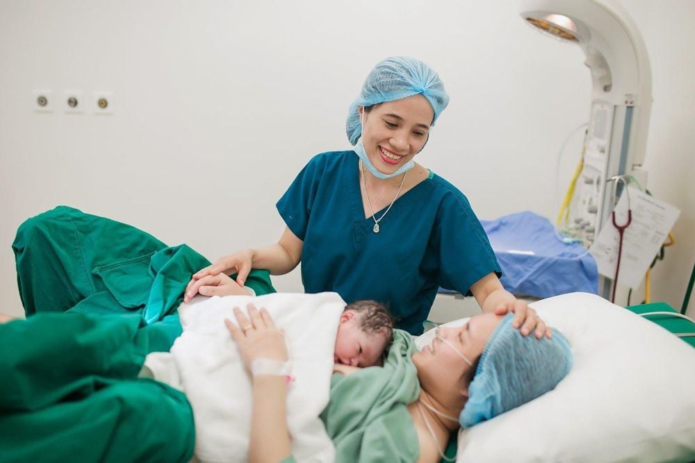
At Vinmec International General Hospital, there is a package maternity service as a solution to help pregnant women feel secure because of the companionship of the medical team throughout the pregnancy. When choosing Maternity Package, pregnant women can:
The pregnancy process is monitored by a team of qualified doctors Regular check-up, early detection of abnormalities Maternity package helps to facilitate the process. birthing process Newborns get comprehensive care
Please dial HOTLINE for more information or register for an appointment HERE. Download MyVinmec app to make appointments faster and to manage your bookings easily.


