This is an automatically translated article.
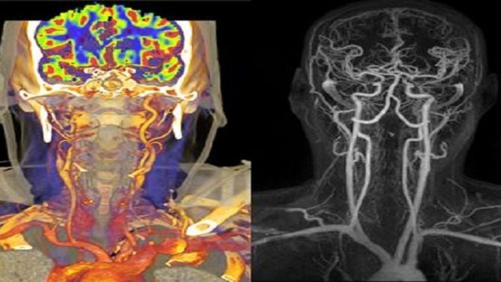
The article is professionally consulted by Master, Doctor Nguyen Viet Thu - Doctor of Radiology and Nuclear Medicine - Department of Diagnostic Imaging and Nuclear Medicine - Vinmec Times City International HospitalCerebral angiography includes computed tomography (CT Angiography - CTA), magnetic resonance imaging (MRA), digital background angiography (DSA), in which CTA is one of the subclinical indications. used to survey the condition of blood vessels feeding the brain, helping to detect narrowing, embolism,... The problem of when to have a CT angiogram of the brain depends on many factors and is being investigated. of great interest in clinical practice today.
1. Learn about CT angiography of the brain
CT cerebral angiography, also known as computed tomography angiography, is a subclinical technique that combines tomography and angiography, to investigate problems related to cerebral blood vessels in a clear way. detail. CTA is the use of a scanner to record images of blood vessels in the brain and create 3D images using specialized software to produce the final image results. Through the results of CT angiography, the doctor can partly orient the diagnosis to diseases such as intracranial stenosis, extracranial stenosis, stroke, aneurysm,... For imaging CT cerebrovascular , the patient needs to be injected with contrast dye into the blood vessels to be able to most accurately visualize the narrowing or non-stenosis of these vessels. This is considered a method that does not cause any invasiveness to the patient, but still brings efficiency and high accuracy, and has great diagnostic value for a number of cerebrovascular diseases. Clinically, in the case of a patient admitted to the hospital because of a cerebral hemorrhage, especially a subarachnoid hemorrhage, a CT angiogram of the brain is indispensable, because the cause as well as the location of the hemorrhage can be found. patient. In addition, CT angiography also contributes to early diagnosis and warning of cerebrovascular stenosis, occlusion or aneurysm even when the patient does not have any clinical symptoms, helping to minimize the risk of complications. from this group of diseases.
2. The benefits and limitations of CT angiography
2.1 . Benefit:
CT angiography of the brain can eliminate the need for surgery. If surgery is still needed, it can be done more precisely. CT angiography is fast, noninvasive, and may have fewer complications than DSA background digitized angiography. Provides more precise anatomical details than other cerebral angiography methods such as DSA and MRA. For CT Angioplasty, no sedation or general anesthesia is required.
2.2 . Limit:
Practically any method will have some limitations that patients need to be aware of and CT angiography is not an exception. The risks that the patient may have when having a CT angiogram of the brain are:Radiation contamination, affecting many organ systems in the patient's body, However, the benefits of an accurate diagnosis exceed far from risk. Allergy to contrast agent. However, the risk of serious allergic reactions to iodinated contrast agents is rare and Vinmec hospitals are well equipped to deal with them. May affect the fetus if the patient is a pregnant woman, so it is necessary to provide full information about yourself and pregnancy status when asked by the doctor. Women who are breast-feeding should not breastfeed for 24 to 48 hours after contrast injection. However, many studies have shown that the amount of contrast agent absorbed by infants during breastfeeding is extremely low. Affects health if the patient is suffering from kidney disease or severe diabetes.

3. When is a CT angiogram of the brain needed?
Some indications for CT angiography are:Patients who frequently present with headache symptoms and are suspected of having some cerebrovascular problems should need a CT angiogram to investigate cerebral circulation, at the same time, early detection if the patient carries dangerous diseases such as brain tumor, cerebrovascular malformation, cerebrovascular accident,... Diagnosis of cerebral infarction also requires CT angiography. brain to find the location of injury, occlusion or narrowing, so that cerebral vascular intervention can be performed right at the damaged place. Screening for brain diseases such as cerebrovascular stenosis, cerebral vascular occlusion, cerebral aneurysm,... Detecting brain tumors. Some basic preparations that patients need to pay attention to before performing a CT angiogram are:
You should wear loose, comfortable clothes when you come for the scan, and don't eat too solid food before the scan. about 4 hours. Do not bring metal objects in the CT scan area such as jewelry, necklaces, eyeglasses, earrings, dentures and hairpins, etc., which may affect the CT image. Patients on sedation should move reasonably gently so that the imaging is not affected. For female subjects of childbearing age, it is necessary to inform the doctor about the status of pregnancy and lactation. During cerebral angiography, the patient will have an intravenous line placed on the inside of the arm or other convenient location to deliver sedation or contrast material into the body. The scanner will work around the patient's body and emit a special noise, the patient needs to stay in the position all the time performing this technique, otherwise, it may result in poor scan results. accuracy as well as in some cases it is necessary to repeat multiple times to be of value in diagnosis. When the first scan is done (scout view), the patient will be given the right amount of contrast material into the body. At the end of the CT angiogram, the patient is monitored for an additional 30 minutes of allergy risk in the imaging room and can live and eat normally, especially need to drink a lot of water during this time to take the medicine. The contrast is soon eliminated from the body.
CT angiography of the brain is a method of high diagnostic value and is indicated in most of the diseases of the blood vessels of the brain and the brain, especially the ischemic stroke or hemorrhagic stroke. However, it also has some limitations, so patients need to pay attention to some recommendations that doctors give. Currently, CT angiography of the brain is one of the imaging techniques applied by Vinmec International General Hospital to help detect brain diseases for timely intervention. Especially to give the most accurate results, the hospital has invested in the most advanced multi-sequence CT and magnetic resonance imaging machines today to serve the examination process. Along with the application of modern equipment is a team of doctors, technicians, experienced experts at home and abroad, who will directly consult, examine and treat to bring the best health. for the patient.
Please dial HOTLINE for more information or register for an appointment HERE. Download MyVinmec app to make appointments faster and to manage your bookings easily.


