This is an automatically translated article.
Article by Doctor Pham Quoc Thanh - Department of Diagnostic Imaging - Vinmec Hai Phong International General Hospital.
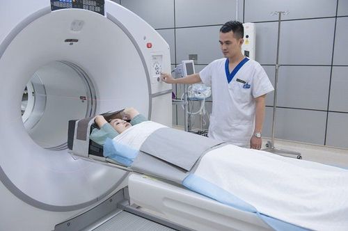
Computed tomography (CT) is an imaging technique. This technique is a machine system that emits many X-rays to scan an area of the body in a cross-section, combined with computer processing to produce 2-D or 3-D images of the part needed. take a shot.
1. What is a CT angiogram?
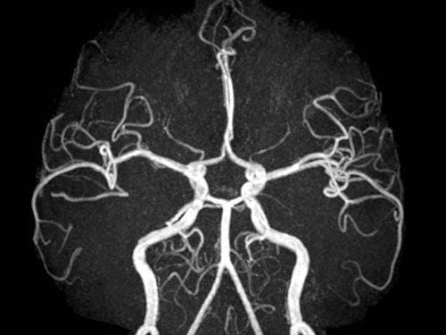
CT angiography is an imaging technique that looks at the blood vessels in the brain. A scanner is used to take pictures of the blood vessels, which then reconstruct a three-dimensional (3D) image using specialized software on a computer.
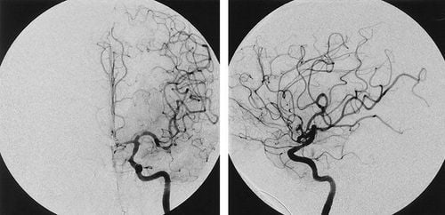
For most emergency cases, CT scan is usually the first priority method after brain magnetic resonance angiography, percutaneous cerebral angiography because it gives faster results, this is very valuable in assessment of patients with acute stroke.
Based on the images obtained from CT angiography, it is possible to diagnose a number of diseases such as intracranial or extracranial stenosis, the risk of stroke or aneurysm... This is a non-invasive technique, has high accuracy and is very valuable in surveying cerebral blood vessels.
In addition, this method also helps to screen for diseases such as narrowing / occlusion of cerebral blood vessels or cerebral aneurysms even when there are no specific symptoms.
2. When is CT angiography indicated?
2.1. Indications: Suspected cases of cerebrovascular abnormalities such as subarachnoid hemorrhage, cerebral parenchymal bleeding, intraventricular bleeding... Cerebral vascular malformations, epilepsy suspected of cerebral vascular malformations Stroke cerebrovascular: Arterial infarction, venous infarction need to evaluate the source of the blood supply to the tumor... Follow up after treatment for cerebrovascular disease. In the case of surgical intervention or after endovascular intervention, it is necessary to require a machine with more than 64 cutting layers to evaluate the noise area of the metal. 2.2. Contraindications In the examination area (cranial) there are many metals that interfere with the image (relative contraindication) Pregnant patients (relative contraindication). Patients with contraindications to the use of iodinated contrast agents.

3. Notes before CT cerebral angiography?
The patient should follow the instructions of the radiologist. Depending on your specific situation, the staff will give specific instructions, the preparation operations are the same, specifically as follows:
4 hours before the CT scan, do not eat solid food. With proper dressing instructions, personal items will be locked with a separate locker and you will hold the key. Do not wear rings, chains, ... metal jewelry including hairpins or dentures. If the subject is using sedatives, a reasonable means of transportation must be arranged. The patient fills in the consent form completely so that the diagnostician can understand your medical history.
4. Steps to perform a CT angiogram of the brain
Step 1: You will go to the shooting room. The technician will help you lie on the CT table. The technician will explain the problems that occur during the shooting.
Step 2: An intravenous line will be made in your hand.
Step 3: During the scan, which lasts less than ten minutes, you will hear buzzing and noises as the CT scanner rotates around your body. It is extremely important to lie still during the CT scan to avoid blurring of the image.
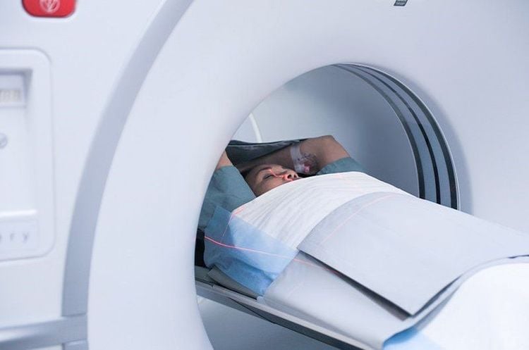
Step 4: The technicians will take pictures, you will see the table is moving out, put in. A small amount of contrast dye is then put into your body through an IV line to gauge the time it takes for the drug to reach the brain. The auto-injector then injects a larger amount of contrast material into your body. You may feel a burning sensation or metallic taste in your mouth as the medicine is injected into your body. The machine will take a snapshot of all images and the auto-injector will inject contrast as needed.
Step 5: Finish shooting. You are allowed to lie down and monitor after the scan. The intravenous line will then be removed.
Step 6: You will change back into your clothes and be able to resume normal activities. You should drink water for an extra day for faster elimination of the drug from your body.
The technicians will not be in the room during the shoot, but you can talk to them through the intercom.
5. CT scan of cerebral blood vessels:
Is an important test in the diagnosis of vascular diseases located in the skull to give the earliest and most accurate diagnosis. Therefore, it is important to seek medical attention immediately, as soon as possible when vascular lesions are suspected.Computed tomography is one of the imaging techniques applied by Vinmec International General Hospital, from which to detect brain diseases, cerebrovascular malformations,...for early diagnosis. In order to improve the diagnostic results, Vinmec currently has a 640 series CT scanner system of Hitachi + the accompanying 3d circuit-building software system, for dark images. best. Vinmec has been and is continuing to invest in modern CT, X-ray, and MRI machines,... in combination with medical services for examination, treatment and early screening of cancers. With a team of highly qualified and experienced medical doctors, direct consultation, examination and treatment will bring optimal treatment results to customers.
Please dial HOTLINE for more information or register for an appointment HERE. Download MyVinmec app to make appointments faster and to manage your bookings easily.


