This is an automatically translated article.
The article was professionally consulted by resident Doctor Nguyen Quynh Giang - Department of Diagnostic Imaging and Nuclear Medicine - Vinmec Times City International Hospital.Ultrasound is a noninvasive procedure that uses sound waves, not radiation, to evaluate the structure of organs in the body. Ultrasound is a convenient and easily accessible tool for rapid examination. So what is ultrasound used for?
1. Purpose of Ultrasound
Ultrasound is a technique that uses high-frequency sound waves to capture images of organs, organ systems, and internal organs without using other invasive methods such as surgery. Ultrasound is increasingly developed and not only records images of tissues, ultrasound can now describe the movement of organs or blood vessels in the body.Unlike X-rays or computed tomography, ultrasound does not use radiation, so it is safe for the body, especially in the case of fetal ultrasound.
Ultrasound imaging is a valuable diagnostic tool in medicine. Depending on the doctor's instructions, ultrasound can have the following purposes:
● Abdominal ultrasound: The purpose of abdominal ultrasound is to observe tissues and organs in this area such as the liver, pancreas, spleen... to find out the unusual signs.
● Bone ultrasound: Ultrasound to evaluate bone thinning helps doctors give advice to limit the patient's fracture
● Breast ultrasound: Observe and screen for early signs of cancer breast.
● Doppler ultrasound: Calculates blood flow and speed in blood vessels, detects blood clots that obstruct blood flow and other heart-related diseases.
Echocardiography : Observe and detect abnormalities of the heart

Siêu âm tim giúp bác sĩ phát hiện bất thường của tim
● Biopsy : Ultrasound can help technicians get the exact cells from the tissue that needs to be biopsied.
2. Ultrasound technique
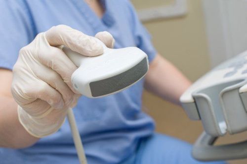
The ultrasound technique is relatively simple:First, before performing the ultrasound, the doctor or technician will ask the patient to take off the jewelry and a part of the clothes where the ultrasound transducer is located. towards then lie on the hospital bed
● For conventional ultrasound techniques such as abdominal ultrasound, the technician will gently apply a layer of gel to the patient's abdomen. This gel layer both allows the ultrasound probe to move easily and prevents gaps between the transducer and the skin that could affect the results. After adjusting the probe to obtain the clearest image for diagnosis, the patient will be wiped off the gel on the skin, dressed again and instructed to wait for the results.
● For natural transvaginal ultrasound techniques such as vaginal or anal ultrasound, instead of just moving on the skin of the body, the transducer will be gently inserted by the technician into the vaginal or anal opening to Obtain the images required by the doctor. This technique is often used in early pregnancy ultrasound, to detect some infections in women or to evaluate the function of the rectum or prostate gland.
In cases where endoscopic ultrasound or cell biopsy is indicated, the ultrasound probes will be attached to the endoscope, through the esophagus into the body to observe the heart, blood vessels or help with the technique. The tablet takes exactly the cells in the area to be biopsied.

Hình ảnh mô phỏng kỹ thuật siêu âm đầu dò qua lỗ tự nhiên (âm đạo)
Today, ultrasound has been widely applied in medicine with many different purposes such as observing damage to tissues and organs to make appropriate diagnosis and treatment, taking advantage of ultrasound for restorative therapy. Function and especially fetal ultrasound is one of the most used techniques to help parents monitor their child's development right from the time they are in the womb. In addition, with the advantages of safety, ease of implementation along with a short time and reasonable cost, ultrasound is increasingly developing and becoming an indispensable technique for humans.
Vinmec International General Hospital system uses the most modern generations of color ultrasound machines today in examining and treating patients. In addition, a team of experienced doctors and nurses will greatly assist in the diagnosis and early detection of abnormal signs of the body in order to provide timely treatment.
Doctor Giang has many years of experience in the field of diagnostic imaging, especially in the field of multi-slice computed tomography, magnetic resonance. Currently, the doctor is working at the Department of Diagnostic Imaging and Nuclear Medicine - Vinmec Times City International General Hospital.
To register for examination and treatment at Vinmec International General Hospital, you can contact the nationwide Vinmec Health System Hotline, or register online HERE.
Source: fda.gov & mayoclinic