This is an automatically translated article.
Tumor is a rare condition and subungual cystic tumor is the most common condition. They can appear in other locations of the outer finger, including the lobe area. So what is vascular tumor and how to recognize and treat this disease?
1. What is U coil circuit?
The coil is an anatomical structure located just below the skin, it is present in many different places on our body, as well as in organs, but the most common is still on the fingertips and toes.
This is a complex structure in which mainly catheters connect directly from the artery to the vein without going through the capillary network. In addition, this system has cell types and nerve endings that accompany the blood vessels. These coils play an important role in regulating body temperature.
Tumor is a tumor growing on the basis of the structure of the coil, first discovered in 1812 and then mentioned by many authors. But it was not until 1924 that the disease was fully described by Masson.
Tumor is a rare disease, accounting for less than 2% of soft tissue tumors, the malignancy rate of this tumor is also very low (<1%), accounting for less than 1% of tumors of the hand. Tumors in the tips of the fingers and toes are the most common, but can also occur in other locations or in many locations. Depending on the predominant anatomical component, choroidal neoplasms can be divided into many smaller types.
2. Signs to recognize coil u
The patient's discomforts appear mainly when there is a collision in the finger lobe area during daily activities and work. The pain may radiate up the upper part of the finger, the pain is usually short-lived and reappears quickly when the next stimulus is present.
Patients often present with pain upon mechanical or thermal stimulation. The pain usually comes on quickly, radiating from the tip of the finger, then gradually subsides and goes away on its own, rarely lasting. In the absence of stimulation, the patient has no pain symptoms.
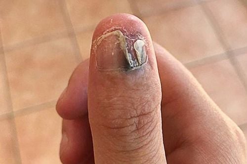
These symptoms appear because the nerve endings are not covered by myelin sheath, so they are stimulated. When the tumor is large, you can see a blue area under the nail. However, coiled tumor located in the lobe position is not clinically observable, direct stimuli will cause symptoms of nerve excitability which is quite obvious but not easy to detect. It is possible to localize the location of the vascular tumor because it is located quite deep.
At an early stage, it is often difficult to diagnose because the lesions are small and the clinical manifestations are not really clear, so this disease is often detected late.

Biểu hiện u cuộn mạch khi kích thước lớn là vùng màu xanh phía dưới móng tay
3. How to diagnose choroid tumor?
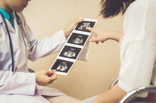
There are two main imaging methods that can be used to confirm the diagnosis of choroid cysts: ultrasound and MRI.
Ultrasound can see a hypoechoic mass under the nail, but it is difficult in some cases such as the tumor is too small or interference due to the patient's fingernails. Ultrasound helps to assess the size and location of the tumor to facilitate surgery. Magnetic resonance has the advantage of helping to accurately evaluate choroidal tumors, but they are expensive. In addition, in some forms of choroidal neoplasms, it is difficult to detect because it is homogenous to the patient's fingernails. The typical image of a choroid on MRI is co-intensity with the patient's fingernails on T1 and hyperintensity on T2. Besides, magnetic resonance imaging and ultrasound, especially Doppler ultrasound, are valuable in differential diagnosis for some other tumor lesions under the nail. The MRI film also helps the surgeon choose the right incision to remove the tumor.
4. How to treat choroid tumor?
Once a definite diagnosis of subungual angioma is made, surgical treatment is the only option. The surgical technique of choroidectomy is relatively simple, bringing high efficiency.
The surgery is performed with an incision about 1.5cm towards the outer edge of the third knuckle of the finger, then the doctor will dissect a milky white tumor with a diameter of about 2mm corresponding to the specified location. previously on magnetic resonance film.
Tumor is a rare disease that can be treated with surgery. Therefore, when being diagnosed with a vascular tumor, the patient needs to go to specialized medical facilities for examination and surgical treatment.
With many years of experience in examining and treating musculoskeletal diseases, performing orthopedic surgery, now Vinmec International General Hospital has become one of the health care centers large, capable of examining, screening and treating many diseases in depth. Therefore, if there are signs of vascular tumor, you can go to Vinmec International General Hospital for examination, diagnosis and treatment to help restore health quickly. Musculoskeletal doctors and doctors at the Orthopedic Trauma Center with expertise and experience will combine consultations and implement the most advanced diagnostic methods available today to assess the patient's condition. and specify a specific treatment regimen.
Please dial HOTLINE for more information or register for an appointment HERE. Download MyVinmec app to make appointments faster and to manage your bookings easily.